Structural Basis of the Heterodimer Formation between Cell Shape-Determining Proteins Csd1 and Csd2 from Helicobacter pylori
An, D.R., Im, H.N., Jang, J.Y., Kim, H.S., Kim, J., Yoon, H.J., Hesek, D., Lee, M., Mobashery, S., Kim, S.J., Suh, S.W.(2016) PLoS One 11: e0164243-e0164243
- PubMed: 27711177
- DOI: https://doi.org/10.1371/journal.pone.0164243
- Primary Citation of Related Structures:
5J1K, 5J1L, 5J1M - PubMed Abstract:
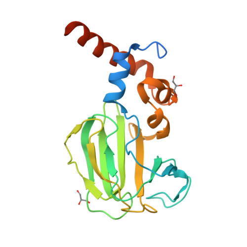
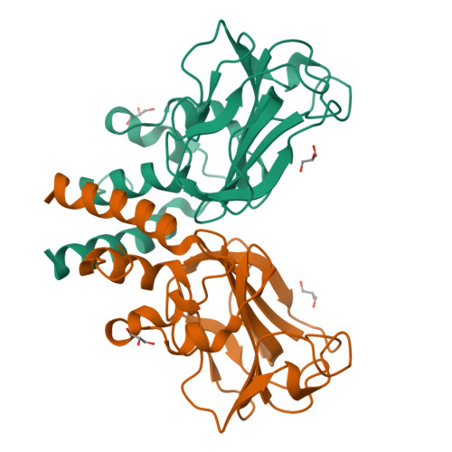
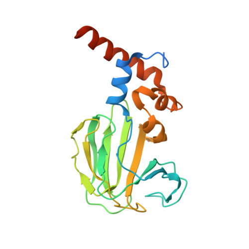
Colonization of the human gastric mucosa by Helicobacter pylori requires its high motility, which depends on the helical cell shape. In H. pylori, several genes (csd1, csd2, csd3/hdpA, ccmA, csd4, csd5, and csd6) play key roles in determining the cell shape by alteration of cross-linking or by trimming of peptidoglycan stem peptides. H. pylori Csd1, Csd2, and Csd3/HdpA are M23B metallopeptidase family members and may act as d,d-endopeptidases to cleave the d-Ala4-mDAP3 peptide bond of cross-linked dimer muropeptides. Csd3 functions also as the d,d-carboxypeptidase to cleave the d-Ala4-d-Ala5 bond of the muramyl pentapeptide. To provide a basis for understanding molecular functions of Csd1 and Csd2, we have carried out their structural characterizations. We have discovered that (i) Csd2 exists in monomer-dimer equilibrium and (ii) Csd1 and Csd2 form a heterodimer. We have determined crystal structures of the Csd2121-308 homodimer and the heterodimer between Csd1125-312 and Csd2121-308. Overall structures of Csd1125-312 and Csd2121-308 monomers are similar to each other, consisting of a helical domain and a LytM domain. The helical domains of both Csd1 and Csd2 play a key role in the formation of homodimers or heterodimers. The Csd1 LytM domain contains a catalytic site with a Zn2+ ion, which is coordinated by three conserved ligands and two water molecules, whereas the Csd2 LytM domain has incomplete metal ligands and no metal ion is bound. Structural knowledge of these proteins sheds light on the events that regulate the cell wall in H. pylori.
Organizational Affiliation:
Department of Biophysics and Chemical Biology, College of Natural Sciences, Seoul National University, Seoul, Korea.


