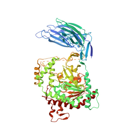
5IXC

Human GIVD cytosolic phospholipase A2 in complex with Methyl gamma-Linolenyl Fluorophosphonate


Experimental Data Snapshot

  • Method: X-RAY DIFFRACTION
  • Resolution: 2.65 Å
  • R-Value Free: 0.251 
  • R-Value Work: 0.237 
  • R-Value Observed: 0.237 

Starting Model: experimental
View more details

wwPDB Validation   3D Report Full Report


Ligand Structure Quality Assessment 


This is version 1.3 of the entry. See complete history


Literature

Structure of Human GIVD Cytosolic Phospholipase A2 Reveals Insights into Substrate Recognition.

Wang, H.Klein, M.G.Snell, G.Lane, W.Zou, H.Levin, I.Li, K.Sang, B.C.

(2016) J Mol Biol 428: 2769-2779

  • DOI: https://doi.org/10.1016/j.jmb.2016.05.012
  • Primary Citation of Related Structures:  
    5IXC, 5IZ5, 5IZR

  • PubMed Abstract: 

    Cytosolic phospholipases A2 (cPLA2s) consist of a family of calcium-sensitive enzymes that function to generate lipid second messengers through hydrolysis of membrane-associated glycerophospholipids. The GIVD cPLA2 (cPLA2δ) is a potential drug target for developing a selective therapeutic agent for the treatment of psoriasis. Here, we present two X-ray structures of human cPLA2δ, capturing an apo state, and in complex with a substrate-like inhibitor. Comparison of the apo and inhibitor-bound structures reveals conformational changes in a flexible cap that allows the substrate to access the relatively buried active site, providing new insight into the mechanism for substrate recognition. The cPLA2δ structure reveals an unexpected second C2 domain that was previously unrecognized from sequence alignments, placing cPLA2δ into the class of membrane-associated proteins that contain a tandem pair of C2 domains. Furthermore, our structures elucidate novel inter-domain interactions and define three potential calcium-binding sites that are likely important for regulation and activation of enzymatic activity. These findings provide novel insights into the molecular mechanisms governing cPLA2's function in signal transduction.


  • Organizational Affiliation

    Department of Structural Biology, Takeda California, San Diego, CA 92121, USA. Electronic address: wanghui.takeda@gmail.com.


Macromolecules
Find similar proteins by:  (by identity cutoff)  |  3D Structure
Entity ID: 1
MoleculeChains Sequence LengthOrganismDetailsImage
Cytosolic phospholipase A2 delta
A, B
814Homo sapiensMutation(s): 0 
Gene Names: PLA2G4D
EC: 3.1.1.4
Membrane Entity: Yes 
UniProt & NIH Common Fund Data Resources
Find proteins for Q86XP0 (Homo sapiens)
Explore Q86XP0 
Go to UniProtKB:  Q86XP0
PHAROS:  Q86XP0
GTEx:  ENSG00000159337 
Entity Groups  
Sequence Clusters30% Identity50% Identity70% Identity90% Identity95% Identity100% Identity
UniProt GroupQ86XP0
Sequence Annotations
Expand
  • Reference Sequence
Experimental Data & Validation

Experimental Data

  • Method: X-RAY DIFFRACTION
  • Resolution: 2.65 Å
  • R-Value Free: 0.251 
  • R-Value Work: 0.237 
  • R-Value Observed: 0.237 
  • Space Group: P 21 21 21
Unit Cell:
Length ( Å )Angle ( ˚ )
a = 100.567α = 90
b = 111.379β = 90
c = 157.645γ = 90
Software Package:
Software NamePurpose
REFMACrefinement
HKL-2000data reduction
HKL-2000data scaling
PHASERphasing

Structure Validation

View Full Validation Report


Ligand Structure Quality Assessment 


Entry History 

Deposition Data

Revision History  (Full details and data files)

  • Version 1.0: 2016-06-08
    Type: Initial release
  • Version 1.1: 2016-06-22
    Changes: Database references
  • Version 1.2: 2023-09-27
    Changes: Data collection, Database references, Derived calculations, Refinement description
  • Version 1.3: 2024-10-30
    Changes: Structure summary