5IV3 | pdb_00005iv3

Crystal structure of human soluble adenylyl cyclase in complex with alpha,beta-methyleneadenosine-5'-triphosphate and the allosteric inhibitor LRE1


Experimental Data Snapshot

  • Method: X-RAY DIFFRACTION
  • Resolution: 1.86 Å
  • R-Value Free: 
    0.223 (Depositor), 0.225 (DCC) 
  • R-Value Work: 
    0.176 (Depositor), 0.184 (DCC) 
  • R-Value Observed: 
    0.178 (Depositor) 

Starting Model: experimental
View more details

wwPDB Validation 3D Report Full Report

Validation slider image for 5IV3

Ligand Structure Quality Assessment 


This is version 1.3 of the entry. See complete history

Literature

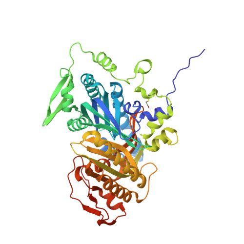
Discovery of LRE1 as a specific and allosteric inhibitor of soluble adenylyl cyclase.

Ramos-Espiritu, L.Kleinboelting, S.Navarrete, F.A.Alvau, A.Visconti, P.E.Valsecchi, F.Starkov, A.Manfredi, G.Buck, H.Adura, C.Zippin, J.H.van den Heuvel, J.Glickman, J.F.Steegborn, C.Levin, L.R.Buck, J.

(2016) Nat Chem Biol 12: 838-844

  • DOI: https://doi.org/10.1038/nchembio.2151
  • Primary Citation Related Structures: 
    5IV3, 5IV4

  • PubMed Abstract: 

    The prototypical second messenger cAMP regulates a wide variety of physiological processes. It can simultaneously mediate diverse functions by acting locally in independently regulated microdomains. In mammalian cells, two types of adenylyl cyclase generate cAMP: G-protein-regulated transmembrane adenylyl cyclases and bicarbonate-, calcium- and ATP-regulated soluble adenylyl cyclase (sAC). Because each type of cyclase regulates distinct microdomains, methods to distinguish between them are needed to understand cAMP signaling. We developed a mass-spectrometry-based adenylyl cyclase assay, which we used to identify a new sAC-specific inhibitor, LRE1. LRE1 bound to the bicarbonate activator binding site and inhibited sAC via a unique allosteric mechanism. LRE1 prevented sAC-dependent processes in cellular and physiological systems, and it will facilitate exploration of the therapeutic potential of sAC inhibition.


  • Organizational Affiliation
    • Department of Pharmacology, Weill Cornell Medical College, New York, New York, USA.

Macromolecule Content 

  • Total Structure Weight: 55.69 kDa 
  • Atom Count: 4,005 
  • Modeled Residue Count: 460 
  • Deposited Residue Count: 475 
  • Unique protein chains: 1

Macromolecules

Find similar proteins by:|  3D Structure
Entity ID: 1
MoleculeChains  Sequence LengthOrganismDetailsImage
Adenylate cyclase type 10475Homo sapiensMutation(s): 0 
Gene Names: ADCY10SAC
EC: 4.6.1.1
UniProt & NIH Common Fund Data Resources
Find proteins for Q96PN6 (Homo sapiens)
Explore Q96PN6 
Go to UniProtKB:  Q96PN6
PHAROS:  Q96PN6
GTEx:  ENSG00000143199 
Entity Groups
Sequence Clusters30% Identity50% Identity70% Identity90% Identity95% Identity100% Identity
UniProt GroupQ96PN6
Sequence Annotations
Expand
Reference Sequence

Small Molecules

Ligands 8 Unique
IDChains Name / Formula / InChI Key2D Diagram3D Interactions
APC

Query on APC


Download:Ideal Coordinates CCD File
H [auth A]DIPHOSPHOMETHYLPHOSPHONIC ACID ADENOSYL ESTER
C11 H18 N5 O12 P3
CAWZRIXWFRFUQB-IOSLPCCCSA-N
LRI

Query on LRI


Download:Ideal Coordinates CCD File
G [auth A]6-chloro-N~4~-cyclopropyl-N~4~-[(thiophen-2-yl)methyl]pyrimidine-2,4-diamine
C12 H13 Cl N4 S
PDWZXKSZLRVSEH-UHFFFAOYSA-N
PEG

Query on PEG


Download:Ideal Coordinates CCD File
J [auth A]DI(HYDROXYETHYL)ETHER
C4 H10 O3
MTHSVFCYNBDYFN-UHFFFAOYSA-N
GOL

Query on GOL


Download:Ideal Coordinates CCD File
D [auth A],
E [auth A],
F [auth A]
GLYCEROL
C3 H8 O3
PEDCQBHIVMGVHV-UHFFFAOYSA-N
EDO

Query on EDO


Download:Ideal Coordinates CCD File
K [auth A]1,2-ETHANEDIOL
C2 H6 O2
LYCAIKOWRPUZTN-UHFFFAOYSA-N
ACT

Query on ACT


Download:Ideal Coordinates CCD File
B [auth A],
C [auth A]
ACETATE ION
C2 H3 O2
QTBSBXVTEAMEQO-UHFFFAOYSA-M
CA

Query on CA


Download:Ideal Coordinates CCD File
I [auth A]CALCIUM ION
Ca
BHPQYMZQTOCNFJ-UHFFFAOYSA-N
CL

Query on CL


Download:Ideal Coordinates CCD File
L [auth A]CHLORIDE ION
Cl
VEXZGXHMUGYJMC-UHFFFAOYSA-M
Modified Residues  1 Unique
IDChains TypeFormula2D DiagramParent
CME
Query on CME
A
L-PEPTIDE LINKINGC5 H11 N O3 S2CYS
Binding Affinity Annotations 
IDSourceBinding Affinity
LRI BindingDB:  5IV3 IC50: min: 1500, max: 6300 (nM) from 3 assay(s)

Experimental Data & Validation

Experimental Data

  • Method: X-RAY DIFFRACTION
  • Resolution: 1.86 Å
  • R-Value Free:  0.223 (Depositor), 0.225 (DCC) 
  • R-Value Work:  0.176 (Depositor), 0.184 (DCC) 
  • R-Value Observed: 0.178 (Depositor) 
Space Group: P 63
Unit Cell:
Length ( Å )Angle ( ˚ )
a = 100.032α = 90
b = 100.032β = 90
c = 98.658γ = 120
Software Package:
Software NamePurpose
REFMACrefinement
XDSdata reduction
XSCALEdata scaling
MOLREPphasing

Structure Validation

View Full Validation Report


Ligand Structure Quality Assessment 


Entry History 

& Funding Information

Deposition Data


Funding OrganizationLocationGrant Number
German Research FoundationGermanySTE1701/11

Revision History  (Full details and data files)

  • Version 1.0: 2016-08-17
    Type: Initial release
  • Version 1.1: 2016-09-28
    Changes: Database references
  • Version 1.2: 2024-01-10
    Changes: Author supporting evidence, Data collection, Database references, Derived calculations, Refinement description
  • Version 1.3: 2024-11-13
    Changes: Structure summary