5IU0 | pdb_00005iu0

Rubisco from Arabidopsis thaliana


Experimental Data Snapshot

  • Method: X-RAY DIFFRACTION
  • Resolution: 1.50 Å
  • R-Value Free: 
    0.152 (Depositor), 0.152 (DCC) 
  • R-Value Work: 
    0.136 (Depositor), 0.137 (DCC) 
  • R-Value Observed: 
    0.145 (Depositor) 

Starting Model: experimental
View more details

wwPDB Validation 3D Report Full Report

Validation slider image for 5IU0

Ligand Structure Quality Assessment 


This is version 1.4 of the entry. See complete history

Literature

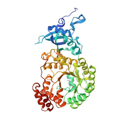
Structure of Rubisco from Arabidopsis thaliana in complex with 2-carboxyarabinitol-1,5-bisphosphate.

Valegard, K.Hasse, D.Andersson, I.Gunn, L.H.

(2018) Acta Crystallogr D Struct Biol 74: 1-9

  • DOI: https://doi.org/10.1107/S2059798317017132
  • Primary Citation Related Structures: 
    5IU0

  • PubMed Abstract: 

    The crystal structure of ribulose-1,5-bisphosphate carboxylase/oxygenase (Rubisco) from Arabidopsis thaliana is reported at 1.5 Å resolution. In light of the importance of A. thaliana as a model organism for understanding higher plant biology, and the pivotal role of Rubisco in photosynthetic carbon assimilation, there has been a notable absence of an A. thaliana Rubisco crystal structure. A. thaliana Rubisco is an L 8 S 8 hexadecamer comprising eight plastome-encoded catalytic large (L) subunits and eight nuclear-encoded small (S) subunits. A. thaliana produces four distinct small-subunit isoforms (RbcS1A, RbcS1B, RbcS2B and RbcS3B), and this crystal structure provides a snapshot of A. thaliana Rubisco containing the low-abundance RbcS3B small-subunit isoform. Crystals were obtained in the presence of the transition-state analogue 2-carboxy-D-arabinitol-1,5-bisphosphate. A. thaliana Rubisco shares the overall fold characteristic of higher plant Rubiscos, but exhibits an interesting disparity between sequence and structural relatedness to other Rubisco isoforms. These results provide the structural framework to understand A. thaliana Rubisco and the potential catalytic differences that could be conferred by alternative A. thaliana Rubisco small-subunit isoforms.


  • Organizational Affiliation
    • Laboratory of Molecular Biophysics, Department of Cell and Molecular Biology, Uppsala University, Husargatan 3, Box 596, SE-751 24 Uppsala, Sweden.

Macromolecule Content 

  • Total Structure Weight: 148.31 kDa 
  • Atom Count: 10,407 
  • Modeled Residue Count: 1,172 
  • Deposited Residue Count: 1,320 
  • Unique protein chains: 2

Macromolecules

Find similar proteins by:|  3D Structure
Entity ID: 1
MoleculeChains  Sequence LengthOrganismDetailsImage
Ribulose bisphosphate carboxylase large chain
A, B
479Arabidopsis thalianaMutation(s): 0 
EC: 4.1.1.39
UniProt
Find proteins for O03042 (Arabidopsis thaliana)
Explore O03042 
Go to UniProtKB:  O03042
Entity Groups
Sequence Clusters30% Identity50% Identity70% Identity90% Identity95% Identity100% Identity
UniProt GroupO03042
Sequence Annotations
Expand
Reference Sequence
Find similar proteins by:|  3D Structure
Entity ID: 2
MoleculeChains  Sequence LengthOrganismDetailsImage
Ribulose bisphosphate carboxylase small chain 1B, chloroplasticC [auth J],
D [auth I]
181Arabidopsis thalianaMutation(s): 0 
EC: 4.1.1.39
UniProt
Find proteins for P10796 (Arabidopsis thaliana)
Explore P10796 
Go to UniProtKB:  P10796
Entity Groups
Sequence Clusters30% Identity50% Identity70% Identity90% Identity95% Identity100% Identity
UniProt GroupP10796
Sequence Annotations
Expand
Reference Sequence

Small Molecules

Ligands 3 Unique
IDChains Name / Formula / InChI Key2D Diagram3D Interactions
CAP

Query on CAP


Download:Ideal Coordinates CCD File
F [auth A],
P [auth B]
2-CARBOXYARABINITOL-1,5-DIPHOSPHATE
C6 H14 O13 P2
ITHCSGCUQDMYAI-ZMIZWQJLSA-N
EDO

Query on EDO


Download:Ideal Coordinates CCD File
G [auth A]
H [auth A]
I [auth A]
J [auth A]
K [auth A]
G [auth A],
H [auth A],
I [auth A],
J [auth A],
K [auth A],
L [auth A],
M [auth A],
N [auth A],
Q [auth B],
R [auth B],
S [auth B],
T [auth I],
U [auth I]
1,2-ETHANEDIOL
C2 H6 O2
LYCAIKOWRPUZTN-UHFFFAOYSA-N
MG

Query on MG


Download:Ideal Coordinates CCD File
E [auth A],
O [auth B]
MAGNESIUM ION
Mg
JLVVSXFLKOJNIY-UHFFFAOYSA-N
Modified Residues  1 Unique
IDChains TypeFormula2D DiagramParent
KCX
Query on KCX
A, B
L-PEPTIDE LINKINGC7 H14 N2 O4LYS

Experimental Data & Validation

Experimental Data

  • Method: X-RAY DIFFRACTION
  • Resolution: 1.50 Å
  • R-Value Free:  0.152 (Depositor), 0.152 (DCC) 
  • R-Value Work:  0.136 (Depositor), 0.137 (DCC) 
  • R-Value Observed: 0.145 (Depositor) 
Space Group: I 4
Unit Cell:
Length ( Å )Angle ( ˚ )
a = 111.86α = 90
b = 111.86β = 90
c = 197.71γ = 90
Software Package:
Software NamePurpose
PHENIXrefinement
XDSdata reduction
XSCALEdata scaling
PHASERphasing

Structure Validation

View Full Validation Report


Ligand Structure Quality Assessment 


Entry History 

Deposition Data

Revision History  (Full details and data files)

  • Version 1.0: 2017-07-12
    Type: Initial release
  • Version 1.1: 2018-01-03
    Changes: Database references
  • Version 1.2: 2018-02-07
    Changes: Database references
  • Version 1.3: 2019-10-16
    Changes: Data collection
  • Version 1.4: 2024-01-10
    Changes: Data collection, Database references, Derived calculations, Refinement description