5IQO | pdb_00005iqo

Crystal structure of the E. coli type 1 pilus subunit FimG (engineered variant with substitutions Q134E and S138E; N-terminal FimG residues 1-12 truncated) in complex with the donor strand peptide DsF_T4R-T6R-D13N


Experimental Data Snapshot

  • Method: X-RAY DIFFRACTION
  • Resolution: 1.30 Å
  • R-Value Free: 
    0.170 (Depositor), 0.162 (DCC) 
  • R-Value Work: 
    0.141 (Depositor) 
  • R-Value Observed: 
    0.141 (Depositor) 

Starting Model: experimental
View more details

wwPDB Validation 3D Report Full Report

Validation slider image for 5IQO

This is version 1.4 of the entry. See complete history

Literature

Accelerating the Association of the Most Stable Protein-Ligand Complex by More than Two Orders of Magnitude.

Giese, C.Eras, J.Kern, A.Scharer, M.A.Capitani, G.Glockshuber, R.

(2016) Angew Chem Int Ed Engl 55: 9350-9355

  • DOI: https://doi.org/10.1002/anie.201603652
  • Primary Citation Related Structures: 
    5IQM, 5IQN, 5IQO

  • PubMed Abstract: 

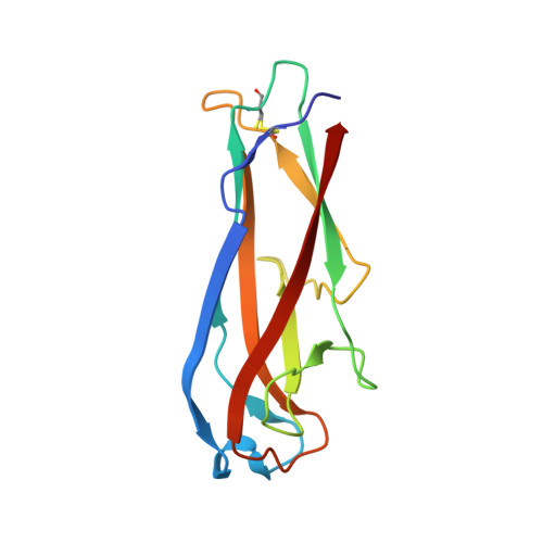
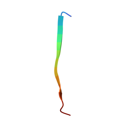
    The complex between the bacterial type 1 pilus subunit FimG and the peptide corresponding to the N-terminal extension (termed donor strand, Ds) of the partner subunit FimF (DsF) shows the strongest reported noncovalent molecular interaction, with a dissociation constant (KD ) of 1.5×10(-20)  m. However, the complex only exhibits a slow association rate of 330 m(-1)  s(-1) that limits technical applications, such as its use in affinity purification. Herein, a structure-based approach was used to design pairs of FimGt (a FimG variant lacking its own N-terminal extension) and DsF variants with enhanced electrostatic surface complementarity. Association of the best mutant FimGt/DsF pairs was accelerated by more than two orders of magnitude, while the dissociation rates and 3D structures of the improved complexes remained essentially unperturbed. A KD  value of 8.8×10(-22)  m was obtained for the best mutant complex, which is the lowest value reported to date for a protein/ligand complex.


  • Organizational Affiliation
    • Department of Biology, ETH Zurich, Otto-Stern-Weg 5, 8093, Zürich, Switzerland.

Macromolecule Content 

  • Total Structure Weight: 31.51 kDa 
  • Atom Count: 2,511 
  • Modeled Residue Count: 293 
  • Deposited Residue Count: 294 
  • Unique protein chains: 2

Macromolecules

Find similar proteins by:|  3D Structure
Entity ID: 1
MoleculeChains  Sequence LengthOrganismDetailsImage
Protein FimG
A, C
132Escherichia coli K-12Mutation(s): 2 
Gene Names: fimGb4319JW4282
UniProt
Find proteins for P08190 (Escherichia coli (strain K12))
Explore P08190 
Go to UniProtKB:  P08190
Entity Groups
Sequence Clusters30% Identity50% Identity70% Identity90% Identity95% Identity100% Identity
UniProt GroupP08190
Sequence Annotations
Expand
Reference Sequence
Find similar proteins by:  Sequence   |   3D Structure  
Entity ID: 2
MoleculeChains  Sequence LengthOrganismDetailsImage
Protein FimF
B, D
15Escherichia coli K-12Mutation(s): 3 
UniProt
Find proteins for P08189 (Escherichia coli (strain K12))
Explore P08189 
Go to UniProtKB:  P08189
Entity Groups
Sequence Clusters30% Identity50% Identity70% Identity90% Identity95% Identity100% Identity
UniProt GroupP08189
Sequence Annotations
Expand
Reference Sequence

Small Molecules

Ligands 3 Unique
IDChains Name / Formula / InChI Key2D Diagram3D Interactions
1PE

Query on 1PE


Download:Ideal Coordinates CCD File
K [auth C]PENTAETHYLENE GLYCOL
C10 H22 O6
JLFNLZLINWHATN-UHFFFAOYSA-N
EDO

Query on EDO


Download:Ideal Coordinates CCD File
I [auth B]1,2-ETHANEDIOL
C2 H6 O2
LYCAIKOWRPUZTN-UHFFFAOYSA-N
CO

Query on CO


Download:Ideal Coordinates CCD File
E [auth A],
F [auth A],
G [auth A],
H [auth A],
J [auth C]
COBALT (II) ION
Co
XLJKHNWPARRRJB-UHFFFAOYSA-N

Experimental Data & Validation

Experimental Data

  • Method: X-RAY DIFFRACTION
  • Resolution: 1.30 Å
  • R-Value Free:  0.170 (Depositor), 0.162 (DCC) 
  • R-Value Work:  0.141 (Depositor) 
  • R-Value Observed: 0.141 (Depositor) 
Space Group: P 1 21 1
Unit Cell:
Length ( Å )Angle ( ˚ )
a = 48.06α = 90
b = 61.85β = 109.12
c = 50.28γ = 90
Software Package:
Software NamePurpose
PHENIXrefinement
XDSdata reduction
XSCALEdata scaling
PHASERphasing

Structure Validation

View Full Validation Report


Entry History 

Deposition Data

Revision History  (Full details and data files)

  • Version 1.0: 2016-07-06
    Type: Initial release
  • Version 1.1: 2016-08-10
    Changes: Database references
  • Version 1.2: 2019-11-20
    Changes: Derived calculations
  • Version 1.3: 2024-01-10
    Changes: Data collection, Database references, Derived calculations, Refinement description
  • Version 1.4: 2024-10-09
    Changes: Structure summary