5INQ

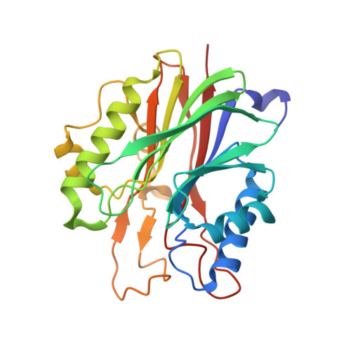
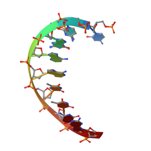
Mouse Tdp2 reaction product (5'-phosphorylated DNA)-Ca2+ complex


Experimental Data Snapshot

  • Method: X-RAY DIFFRACTION
  • Resolution: 1.85 Å
  • R-Value Free: 0.195 
  • R-Value Work: 0.158 
  • R-Value Observed: 0.159 

Starting Model: experimental
View more details

wwPDB Validation   3D Report Full Report


This is version 1.4 of the entry. See complete history


Literature

Reversal of DNA damage induced Topoisomerase 2 DNA-protein crosslinks by Tdp2.

Schellenberg, M.J.Perera, L.Strom, C.N.Waters, C.A.Monian, B.Appel, C.D.Vilas, C.K.Williams, J.G.Ramsden, D.A.Williams, R.S.

(2016) Nucleic Acids Res 44: 3829-3844

  • DOI: https://doi.org/10.1093/nar/gkw228
  • Primary Citation of Related Structures:  
    5HT2, 5INK, 5INL, 5INM, 5INN, 5INO, 5INP, 5INQ

  • PubMed Abstract: 

    Mammalian Tyrosyl-DNA phosphodiesterase 2 (Tdp2) reverses Topoisomerase 2 (Top2) DNA-protein crosslinks triggered by Top2 engagement of DNA damage or poisoning by anticancer drugs. Tdp2 deficiencies are linked to neurological disease and cellular sensitivity to Top2 poisons. Herein, we report X-ray crystal structures of ligand-free Tdp2 and Tdp2-DNA complexes with alkylated and abasic DNA that unveil a dynamic Tdp2 active site lid and deep substrate binding trench well-suited for engaging the diverse DNA damage triggers of abortive Top2 reactions. Modeling of a proposed Tdp2 reaction coordinate, combined with mutagenesis and biochemical studies support a single Mg(2+)-ion mechanism assisted by a phosphotyrosyl-arginine cation-π interface. We further identify a Tdp2 active site SNP that ablates Tdp2 Mg(2+) binding and catalytic activity, impairs Tdp2 mediated NHEJ of tyrosine blocked termini, and renders cells sensitive to the anticancer agent etoposide. Collectively, our results provide a structural mechanism for Tdp2 engagement of heterogeneous DNA damage that causes Top2 poisoning, and indicate that evaluation of Tdp2 status may be an important personalized medicine biomarker informing on individual sensitivities to chemotherapeutic Top2 poisons.


  • Organizational Affiliation

    Genome Integrity and Structural Biology Laboratory, National Institute of Environmental Health Sciences, US National Institutes of Health, Department of Health and Human Services, Research Triangle Park, NC 27709, USA.


Macromolecules

Find similar proteins by:  (by identity cutoff)  |  3D Structure
Entity ID: 1
MoleculeChains Sequence LengthOrganismDetailsImage
Tyrosyl-DNA phosphodiesterase 2
A, B
256Mus musculusMutation(s): 0 
Gene Names: Tdp2Ttrap
EC: 3.1.4
UniProt
Find proteins for Q9JJX7 (Mus musculus)
Explore Q9JJX7 
Go to UniProtKB:  Q9JJX7
Entity Groups  
Sequence Clusters30% Identity50% Identity70% Identity90% Identity95% Identity100% Identity
UniProt GroupQ9JJX7
Sequence Annotations
Expand
  • Reference Sequence

Find similar nucleic acids by:  Sequence   |   3D Structure  

Entity ID: 2
MoleculeChains LengthOrganismImage
DNA (5'-D(P*CP*CP*GP*AP*AP*TP*TP*CP*G)-3')
C, D
9Homo sapiens
Sequence Annotations
Expand
  • Reference Sequence
Small Molecules
Ligands 4 Unique
IDChains Name / Formula / InChI Key2D Diagram3D Interactions
EPE
Query on EPE

Download Ideal Coordinates CCD File 
H [auth B]4-(2-HYDROXYETHYL)-1-PIPERAZINE ETHANESULFONIC ACID
C8 H18 N2 O4 S
JKMHFZQWWAIEOD-UHFFFAOYSA-N
PEG
Query on PEG

Download Ideal Coordinates CCD File 
F [auth A]DI(HYDROXYETHYL)ETHER
C4 H10 O3
MTHSVFCYNBDYFN-UHFFFAOYSA-N
GOL
Query on GOL

Download Ideal Coordinates CCD File 
I [auth B]GLYCEROL
C3 H8 O3
PEDCQBHIVMGVHV-UHFFFAOYSA-N
CA
Query on CA

Download Ideal Coordinates CCD File 
E [auth A],
G [auth B]
CALCIUM ION
Ca
BHPQYMZQTOCNFJ-UHFFFAOYSA-N
Experimental Data & Validation

Experimental Data

  • Method: X-RAY DIFFRACTION
  • Resolution: 1.85 Å
  • R-Value Free: 0.195 
  • R-Value Work: 0.158 
  • R-Value Observed: 0.159 
  • Space Group: P 21 21 21
Unit Cell:
Length ( Å )Angle ( ˚ )
a = 54.702α = 90
b = 67.504β = 90
c = 167.529γ = 90
Software Package:
Software NamePurpose
SCALEPACKdata scaling
PHENIXrefinement
PDB_EXTRACTdata extraction
DENZOdata reduction
PHENIXphasing

Structure Validation

View Full Validation Report


Entry History & Funding Information

Deposition Data


Funding OrganizationLocationGrant Number
National Institutes of Health/National Institute of Environmental Health Sciences (NIH/NIEHS)United States1Z01ES102765

Revision History  (Full details and data files)

  • Version 1.0: 2016-04-27
    Type: Initial release
  • Version 1.1: 2016-05-18
    Changes: Database references
  • Version 1.2: 2017-09-20
    Changes: Author supporting evidence, Database references, Derived calculations
  • Version 1.3: 2019-12-18
    Changes: Author supporting evidence
  • Version 1.4: 2023-09-27
    Changes: Data collection, Database references, Derived calculations, Refinement description