5IMW | pdb_00005imw

Trapped Toxin


Experimental Data Snapshot

  • Method: X-RAY DIFFRACTION
  • Resolution: 2.89 Å
  • R-Value Free: 
    0.295 (Depositor), 0.308 (DCC) 
  • R-Value Work: 
    0.258 (Depositor), 0.269 (DCC) 
  • R-Value Observed: 
    0.260 (Depositor) 

Starting Model: experimental
View more details

wwPDB Validation 3D Report Full Report

Validation slider image for 5IMW

This is version 1.3 of the entry. See complete history

Literature

Structural Basis for Receptor Recognition by the Human CD59-Responsive Cholesterol-Dependent Cytolysins.

Lawrence, S.L.Gorman, M.A.Feil, S.C.Mulhern, T.D.Kuiper, M.J.Ratner, A.J.Tweten, R.K.Morton, C.J.Parker, M.W.

(2016) Structure 24: 1488-1498

  • DOI: https://doi.org/10.1016/j.str.2016.06.017
  • Primary Citation Related Structures: 
    5IMT, 5IMW, 5IMY

  • PubMed Abstract: 

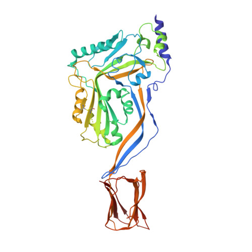
    Cholesterol-dependent cytolysins (CDCs) are a family of pore-forming toxins that punch holes in the outer membrane of eukaryotic cells. Cholesterol serves as the receptor, but a subclass of CDCs first binds to human CD59. Here we describe the crystal structures of vaginolysin and intermedilysin complexed to CD59. These studies, together with small-angle X-ray scattering, reveal that CD59 binds to each at different, though overlapping, sites, consistent with molecular dynamics simulations and binding studies. The CDC consensus undecapeptide motif, which for the CD59-responsive CDCs has a proline instead of a tryptophan in the motif, adopts a strikingly different conformation between the structures; our data suggest that the proline acts as a selectivity switch to ensure CD59-dependent CDCs bind their protein receptor first in preference to cholesterol. The structural data suggest a detailed model of how these water-soluble toxins assemble as prepores on the cell surface.


  • Organizational Affiliation
    • ACRF Rational Drug Discovery Centre, St. Vincent's Institute of Medical Research, Fitzroy, VIC 3065, Australia.

Macromolecule Content 

  • Total Structure Weight: 103.77 kDa 
  • Atom Count: 7,113 
  • Modeled Residue Count: 918 
  • Deposited Residue Count: 942 
  • Unique protein chains: 1

Macromolecules

Find similar proteins by:|  3D Structure
Entity ID: 1
MoleculeChains  Sequence LengthOrganismDetailsImage
Intermedilysin
A, B
471Streptococcus intermediusMutation(s): 0 
Gene Names: ily
Membrane Entity: Yes 
UniProt
Find proteins for Q9LCB8 (Streptococcus intermedius)
Explore Q9LCB8 
Go to UniProtKB:  Q9LCB8
Entity Groups
Sequence Clusters30% Identity50% Identity70% Identity90% Identity95% Identity100% Identity
UniProt GroupQ9LCB8
Sequence Annotations
Expand
Reference Sequence

Experimental Data & Validation

Experimental Data

  • Method: X-RAY DIFFRACTION
  • Resolution: 2.89 Å
  • R-Value Free:  0.295 (Depositor), 0.308 (DCC) 
  • R-Value Work:  0.258 (Depositor), 0.269 (DCC) 
  • R-Value Observed: 0.260 (Depositor) 
Space Group: P 21 2 21
Unit Cell:
Length ( Å )Angle ( ˚ )
a = 84.465α = 90
b = 101.565β = 90
c = 175.735γ = 90
Software Package:
Software NamePurpose
PHENIXrefinement
XSCALEdata scaling
PHASERphasing
PDB_EXTRACTdata extraction
XDSdata reduction

Structure Validation

View Full Validation Report


Entry History 

Deposition Data

Revision History  (Full details and data files)

  • Version 1.0: 2016-08-24
    Type: Initial release
  • Version 1.1: 2016-09-14
    Changes: Database references
  • Version 1.2: 2023-09-27
    Changes: Data collection, Database references, Derived calculations, Refinement description
  • Version 1.3: 2024-10-23
    Changes: Structure summary