5ILV | pdb_00005ilv

Uninhibited ETV5


Experimental Data Snapshot

  • Method: X-RAY DIFFRACTION
  • Resolution: 1.80 Å
  • R-Value Free: 
    0.234 (Depositor), 0.237 (DCC) 
  • R-Value Work: 
    0.186 (Depositor), 0.187 (DCC) 
  • R-Value Observed: 
    0.189 (Depositor) 

wwPDB Validation 3D Report Full Report

Validation slider image for 5ILV

This is version 1.4 of the entry. See complete history

Literature

Structured and disordered regions cooperatively mediate DNA-binding autoinhibition of ETS factors ETV1, ETV4 and ETV5.

Currie, S.L.Lau, D.K.W.Doane, J.J.Whitby, F.G.Okon, M.McIntosh, L.P.Graves, B.J.

(2017) Nucleic Acids Res 45: 2223-2241

  • DOI: https://doi.org/10.1093/nar/gkx068
  • Primary Citation Related Structures: 
    5ILS, 5ILU, 5ILV

  • PubMed Abstract: 

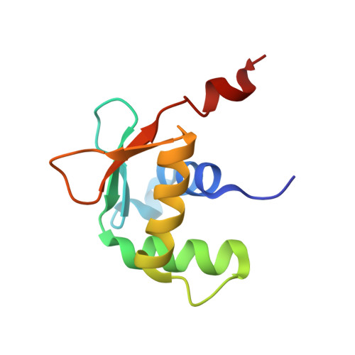
    Autoinhibition enables spatial and temporal regulation of cellular processes by coupling protein activity to surrounding conditions, often via protein partnerships or signaling pathways. We report the molecular basis of DNA-binding autoinhibition of ETS transcription factors ETV1, ETV4 and ETV5, which are often overexpressed in prostate cancer. Inhibitory elements that cooperate to repress DNA binding were identified in regions N- and C-terminal of the ETS domain. Crystal structures of these three factors revealed an α-helix in the C-terminal inhibitory domain that packs against the ETS domain and perturbs the conformation of its DNA-recognition helix. Nuclear magnetic resonance spectroscopy demonstrated that the N-terminal inhibitory domain (NID) is intrinsically disordered, yet utilizes transient intramolecular interactions with the DNA-recognition helix of the ETS domain to mediate autoinhibition. Acetylation of selected lysines within the NID activates DNA binding. This investigation revealed a distinctive mechanism for DNA-binding autoinhibition in the ETV1/4/5 subfamily involving a network of intramolecular interactions not present in other ETS factors. These distinguishing inhibitory elements provide a platform through which cellular triggers, such as protein-protein interactions or post-translational modifications, may specifically regulate the function of these oncogenic proteins.


  • Organizational Affiliation
    • Department of Oncological Sciences, University of Utah, Salt Lake City, UT 84112-5550, USA.

Macromolecule Content 

  • Total Structure Weight: 10.88 kDa 
  • Atom Count: 883 
  • Modeled Residue Count: 92 
  • Deposited Residue Count: 92 
  • Unique protein chains: 1

Macromolecules

Find similar proteins by:|  3D Structure
Entity ID: 1
MoleculeChains  Sequence LengthOrganismDetailsImage
ETS translocation variant 592Homo sapiensMutation(s): 0 
Gene Names: ETV5ERM
UniProt & NIH Common Fund Data Resources
Find proteins for P41161 (Homo sapiens)
Explore P41161 
Go to UniProtKB:  P41161
PHAROS:  P41161
GTEx:  ENSG00000244405 
Entity Groups
Sequence Clusters30% Identity50% Identity70% Identity90% Identity95% Identity100% Identity
UniProt GroupP41161
Sequence Annotations
Expand
Reference Sequence

Experimental Data & Validation

Experimental Data

  • Method: X-RAY DIFFRACTION
  • Resolution: 1.80 Å
  • R-Value Free:  0.234 (Depositor), 0.237 (DCC) 
  • R-Value Work:  0.186 (Depositor), 0.187 (DCC) 
  • R-Value Observed: 0.189 (Depositor) 
Space Group: C 2 2 21
Unit Cell:
Length ( Å )Angle ( ˚ )
a = 57.371α = 90
b = 65.611β = 90
c = 52.677γ = 90
Software Package:
Software NamePurpose
HKL-2000data collection
SCALEPACKdata scaling
PHASERphasing
PHENIXrefinement
PDB_EXTRACTdata extraction

Structure Validation

View Full Validation Report


Entry History 

& Funding Information

Deposition Data


Funding OrganizationLocationGrant Number
National Institutes of Health/National Institute of General Medical Sciences (NIH/NIGMS)United StatesR01GM38663

Revision History  (Full details and data files)

  • Version 1.0: 2017-02-22
    Type: Initial release
  • Version 1.1: 2017-05-03
    Changes: Database references
  • Version 1.2: 2017-09-20
    Changes: Author supporting evidence
  • Version 1.3: 2019-12-25
    Changes: Author supporting evidence
  • Version 1.4: 2024-11-13
    Changes: Data collection, Database references, Structure summary