5II3 | pdb_00005ii3

The X-ray structure of the adduct formed in the reaction between hen egg white lysozyme and compound 3, a platin(II) compound containing a O, S bidentate ligand


Experimental Data Snapshot

  • Method: X-RAY DIFFRACTION
  • Resolution: 1.78 Å
  • R-Value Free: 
    0.190 (Depositor), 0.198 (DCC) 
  • R-Value Work: 
    0.153 (Depositor), 0.167 (DCC) 
  • R-Value Observed: 
    0.155 (Depositor) 

Starting Model: experimental
View more details

wwPDB Validation 3D Report Full Report

Validation slider image for 5II3

This is version 1.2 of the entry. See complete history

Literature

Platinum(ii) O,S complexes as potential metallodrugs against Cisplatin resistance.

Hildebrandt, J.Hafner, N.Gorls, H.Kritsch, D.Ferraro, G.Durst, M.Runnebaum, I.B.Merlino, A.Weigand, W.

(2016) Dalton Trans 45: 18876-18891

  • DOI: https://doi.org/10.1039/c6dt01388k
  • Primary Citation Related Structures: 
    5IHG, 5II3, 5ILC, 5ILF

  • PubMed Abstract: 

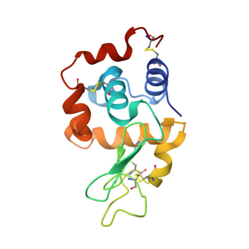
    We report on platinum(ii) complexes with different cinnamic acid derivatives as ligands with cytotoxic activity against Cisplatin resistant ovarian cancer cell line subcultures of SKOV3 and A2780. A typical mechanism of action for platinum(ii) complexes as Cisplatin itself is binding to the DNA and inducing double-strand breaks. We examined the biological behavior of these potential drugs with 9-methylguanine using NMR spectroscopic methods and their DNA damage potential including γH2AX-foci analyses. X-ray diffraction methods have been used to elucidate the molecular structures of the platinum(ii) complexes. Interactions with the model protein lysozyme have been evaluated by different techniques including UV-Vis absorption spectroscopy, fluorescence and X-ray crystallography.


  • Organizational Affiliation
    • Institut für Anorganische und Analytische Chemie Friedrich-Schiller-Universität Jena Humboldtstraße 8, 07743 Jena, Germany. wolfgang.weigand@uni-jena.de.

Macromolecule Content 

  • Total Structure Weight: 14.87 kDa 
  • Atom Count: 1,172 
  • Modeled Residue Count: 129 
  • Deposited Residue Count: 129 
  • Unique protein chains: 1

Macromolecules

Find similar proteins by:|  3D Structure
Entity ID: 1
MoleculeChains  Sequence LengthOrganismDetailsImage
Lysozyme C129Gallus gallusMutation(s): 0 
EC: 3.2.1.17
UniProt
Find proteins for P00698 (Gallus gallus)
Explore P00698 
Go to UniProtKB:  P00698
Entity Groups
Sequence Clusters30% Identity50% Identity70% Identity90% Identity95% Identity100% Identity
UniProt GroupP00698
Sequence Annotations
Expand
Reference Sequence

Small Molecules

Ligands 4 Unique
IDChains Name / Formula / InChI Key2D Diagram3D Interactions
6B6

Query on 6B6


Download:Ideal Coordinates CCD File
C [auth A]platinum(4+) tetrahydroxide
H4 O4 Pt
JTAFSELAEYLDJR-UHFFFAOYSA-J
DMS

Query on DMS


Download:Ideal Coordinates CCD File
B [auth A]DIMETHYL SULFOXIDE
C2 H6 O S
IAZDPXIOMUYVGZ-UHFFFAOYSA-N
CL

Query on CL


Download:Ideal Coordinates CCD File
E [auth A],
F [auth A],
G [auth A],
H [auth A],
I [auth A]
CHLORIDE ION
Cl
VEXZGXHMUGYJMC-UHFFFAOYSA-M
NA

Query on NA


Download:Ideal Coordinates CCD File
D [auth A]SODIUM ION
Na
FKNQFGJONOIPTF-UHFFFAOYSA-N

Experimental Data & Validation

Experimental Data

  • Method: X-RAY DIFFRACTION
  • Resolution: 1.78 Å
  • R-Value Free:  0.190 (Depositor), 0.198 (DCC) 
  • R-Value Work:  0.153 (Depositor), 0.167 (DCC) 
  • R-Value Observed: 0.155 (Depositor) 
Space Group: P 43 21 2
Unit Cell:
Length ( Å )Angle ( ˚ )
a = 78.206α = 90
b = 78.206β = 90
c = 37.005γ = 90
Software Package:
Software NamePurpose
REFMACrefinement
HKL-2000data reduction
HKL-2000data scaling
PHASERphasing

Structure Validation

View Full Validation Report


Entry History 

Deposition Data

Revision History  (Full details and data files)

  • Version 1.0: 2016-12-07
    Type: Initial release
  • Version 1.1: 2024-01-10
    Changes: Data collection, Database references, Refinement description
  • Version 1.2: 2024-11-20
    Changes: Structure summary