5IFG | pdb_00005ifg

Crystal structure of HigA-HigB complex from E. Coli


Experimental Data Snapshot

  • Method: X-RAY DIFFRACTION
  • Resolution: 2.70 Å
  • R-Value Free: 
    0.231 (Depositor), 0.253 (DCC) 
  • R-Value Work: 
    0.199 (Depositor), 0.234 (DCC) 
  • R-Value Observed: 
    0.202 (Depositor) 

wwPDB Validation 3D Report Full Report

Validation slider image for 5IFG

This is version 1.1 of the entry. See complete history

Literature

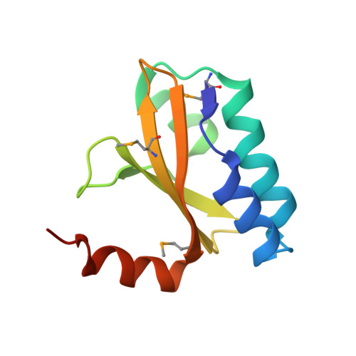
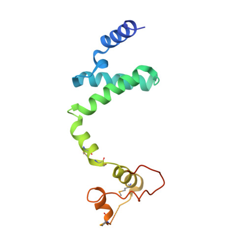
Structural insight into the E. coli HigBA complex

Yang, J.Zhou, K.Liu, P.Dong, Y.Gao, Z.Zhang, J.Liu, Q.

(2016) Biochem Biophys Res Commun 478: 1521-1527

  • DOI: https://doi.org/10.1016/j.bbrc.2016.08.131
  • Primary Citation Related Structures: 
    5IFG

  • PubMed Abstract: 

    The toxin-antitoxin system is ubiquitously existed in bacteria and archaea, performing a wide variety of functions modulating cell fitness in response to environmental cues. In this report, we solved the crystal structure of the toxin-antitoxin HigBA complex from E. coli K-12 to 2.7 Å resolution. The crystal structure of the HigBA complex displays a hetero-tetramer (HigBA)2 form comprised by two HigB and two HigA subunits. Each toxin HigB resumes a microbial RNase T1 fold, characteristic of a three antiparallel β-sheet core shielded by a few α-helices at either side. Each antitoxin HigA composed of all α-helices resembles a "C"-shaped clamp nicely encompassing a HigB in the (HigBA)2 complex. Two HigA monomers dimerize at their N-terminal domain. We showed that HigA helix α1 was essential for HigA dimerization and the hetero-tetramer (HigBA)2 formation, but not for a hetero-dimeric HigBA formation. HigA dimerization mediated by helix α1 was dispensable for DNA-binding, as a heterodimeric HigBA complex still bound to the higBA operator in vitro. The HigA C-terminal domain with a helix-turn-helix fold was essential for DNA binding. We also defined two palindromes in higBA operator specifically recognized by HigA and HigBA in vitro.


  • Organizational Affiliation
    • School of Life Sciences, University of Dalian Science and Technology, Dalian, Liaolin Province, 230027, People's Republic of China.

Macromolecule Content 

  • Total Structure Weight: 55.02 kDa 
  • Atom Count: 3,408 
  • Modeled Residue Count: 435 
  • Deposited Residue Count: 484 
  • Unique protein chains: 2

Macromolecules

Find similar proteins by:|  3D Structure
Entity ID: 1
MoleculeChains  Sequence LengthOrganismDetailsImage
mRNA interferase HigB
A, C
104Escherichia coli K-12Mutation(s): 0 
Gene Names: higBygjNb3083JW3054
EC: 3.1
UniProt
Find proteins for P64578 (Escherichia coli (strain K12))
Explore P64578 
Go to UniProtKB:  P64578
Entity Groups
Sequence Clusters30% Identity50% Identity70% Identity90% Identity95% Identity100% Identity
UniProt GroupP64578
Sequence Annotations
Expand
Reference Sequence
Find similar proteins by:|  3D Structure
Entity ID: 2
MoleculeChains  Sequence LengthOrganismDetailsImage
Antitoxin HigA
B, D
138Escherichia coli K-12Mutation(s): 0 
Gene Names: higAygjMb3082JW3053
UniProt
Find proteins for P67701 (Escherichia coli (strain K12))
Explore P67701 
Go to UniProtKB:  P67701
Entity Groups
Sequence Clusters30% Identity50% Identity70% Identity90% Identity95% Identity100% Identity
UniProt GroupP67701
Sequence Annotations
Expand
Reference Sequence

Small Molecules

Modified Residues  1 Unique
IDChains TypeFormula2D DiagramParent
MSE
Query on MSE
A, C
L-PEPTIDE LINKINGC5 H11 N O2 SeMET

Experimental Data & Validation

Experimental Data

  • Method: X-RAY DIFFRACTION
  • Resolution: 2.70 Å
  • R-Value Free:  0.231 (Depositor), 0.253 (DCC) 
  • R-Value Work:  0.199 (Depositor), 0.234 (DCC) 
  • R-Value Observed: 0.202 (Depositor) 
Space Group: P 31 2 1
Unit Cell:
Length ( Å )Angle ( ˚ )
a = 77.924α = 90
b = 77.924β = 90
c = 180.634γ = 120
Software Package:
Software NamePurpose
PHENIXrefinement
HKL-2000data reduction
HKL-2000data scaling
PHENIXphasing

Structure Validation

View Full Validation Report


Entry History 

Deposition Data

Revision History  (Full details and data files)

  • Version 1.0: 2017-03-01
    Type: Initial release
  • Version 1.1: 2024-10-16
    Changes: Data collection, Database references, Structure summary