5ID7 | pdb_00005id7

Crystal structure of human serum albumin in complex with phosphorodithioate derivative of myristoyl cyclic phosphatidic acid (cPA)


Experimental Data Snapshot

  • Method: X-RAY DIFFRACTION
  • Resolution: 2.26 Å
  • R-Value Free: 
    0.253 (Depositor), 0.249 (DCC) 
  • R-Value Work: 
    0.206 (Depositor), 0.210 (DCC) 
  • R-Value Observed: 
    0.207 (Depositor) 

Starting Model: experimental
View more details

wwPDB Validation 3D Report Full Report

Validation slider image for 5ID7

Ligand Structure Quality Assessment 


This is version 1.3 of the entry. See complete history

Literature

Structural evidence of the species-dependent albumin binding of the modified cyclic phosphatidic acid with cytotoxic properties.

Sekula, B.Ciesielska, A.Rytczak, P.Koziokiewicz, M.Bujacz, A.

(2016) Biosci Rep 36

  • DOI: https://doi.org/10.1042/BSR20160089
  • Primary Citation Related Structures: 
    5ID7, 5ID9

  • PubMed Abstract: 

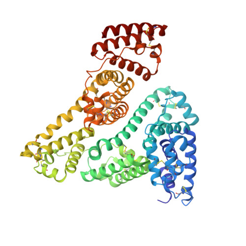
    Cyclic phosphatidic acids (cPAs) are naturally occurring, very active signalling molecules, which are involved in several pathological states, such as cancer, diabetes or obesity. As molecules of highly lipidic character found in the circulatory system, cPAs are bound and transported by the main extracellular lipid binding protein-serum albumin. Here, we present the detailed interactions between human serum albumin (HSA) and equine serum albumin (ESA) with a derivative of cPA, 1-O-myristoyl-sn-glycerol-2,3-cyclic phosphorodithioate (Myr-2S-cPA). Initial selection of the ligand used for the structural study was made by the analysis of the therapeutically promising properties of the sulfur containing analogues of cPA in respect to the unmodified lysophospholipids (LPLs). Substitution of one or two non-bridging oxygen atoms in the phosphate group with one or two sulfur atoms increases the cytotoxic effect of cPAs up to 60% on the human prostate cancer (PC) cells. Myr-2S-cPA reduces cancer cell viability in a dose-dependent manner, with IC50 value of 29.0 μM after 24 h incubation, which is almost 30% lower than IC50 of single substituted phosphorothioate cPA. Although, the structural homology between HSA and ESA is big, their crystal complexes with Myr-2S-cPA demonstrate significantly different mode of binding of this LPL analogue. HSA binds three molecules of Myr-2S-cPA, whereas ESA only one. Moreover, none of the identified Myr-2S-cPA binding sites overlap in both albumins.


  • Organizational Affiliation
    • Institute of Technical Biochemistry, Faculty of Biotechnology and Food Sciences, Lodz University of Technology, Stefanowskiego 4/10, 90-924 Lodz, Poland.

Macromolecule Content 

  • Total Structure Weight: 136.28 kDa 
  • Atom Count: 9,779 
  • Modeled Residue Count: 1,164 
  • Deposited Residue Count: 1,170 
  • Unique protein chains: 1

Macromolecules

Find similar proteins by:|  3D Structure
Entity ID: 1
MoleculeChains  Sequence LengthOrganismDetailsImage
Serum albumin
A, B
585Homo sapiensMutation(s): 0 
UniProt & NIH Common Fund Data Resources
Find proteins for P02768 (Homo sapiens)
Explore P02768 
Go to UniProtKB:  P02768
PHAROS:  P02768
GTEx:  ENSG00000163631 
Entity Groups
Sequence Clusters30% Identity50% Identity70% Identity90% Identity95% Identity100% Identity
UniProt GroupP02768
Sequence Annotations
Expand
Reference Sequence

Small Molecules

Ligands 3 Unique
IDChains Name / Formula / InChI Key2D Diagram3D Interactions
6A4

Query on 6A4


Download:Ideal Coordinates CCD File
C [auth A]
D [auth A]
E [auth A]
J [auth B]
K [auth B]
C [auth A],
D [auth A],
E [auth A],
J [auth B],
K [auth B],
L [auth B]
(4S)-2-sulfanylidene-4-[(tetradecanoyloxy)methyl]-1,3,2lambda~5~-dioxaphospholane-2-thiolate
C17 H32 O4 P S2
PXJKEDDZAWQNJX-INIZCTEOSA-M
PGE

Query on PGE


Download:Ideal Coordinates CCD File
F [auth A],
H [auth A],
M [auth B]
TRIETHYLENE GLYCOL
C6 H14 O4
ZIBGPFATKBEMQZ-UHFFFAOYSA-N
PEG

Query on PEG


Download:Ideal Coordinates CCD File
G [auth A],
I [auth A],
N [auth B]
DI(HYDROXYETHYL)ETHER
C4 H10 O3
MTHSVFCYNBDYFN-UHFFFAOYSA-N

Experimental Data & Validation

Experimental Data

  • Method: X-RAY DIFFRACTION
  • Resolution: 2.26 Å
  • R-Value Free:  0.253 (Depositor), 0.249 (DCC) 
  • R-Value Work:  0.206 (Depositor), 0.210 (DCC) 
  • R-Value Observed: 0.207 (Depositor) 
Space Group: P 1
Unit Cell:
Length ( Å )Angle ( ˚ )
a = 37.895α = 105.46
b = 85.167β = 89.78
c = 96.308γ = 101.34
Software Package:
Software NamePurpose
REFMACrefinement
XDSdata reduction
XSCALEdata scaling

Structure Validation

View Full Validation Report


Ligand Structure Quality Assessment 


Entry History 

& Funding Information

Deposition Data


Funding OrganizationLocationGrant Number
Polish Ministry of Science and Higher EducationPolandN N405 3639 39
National Science Centre PolandPoland2013/11/B/ST5/02271
National Science Centre PolandPoland2014/12/T/ST5/00136

Revision History  (Full details and data files)

  • Version 1.0: 2016-05-11
    Type: Initial release
  • Version 1.1: 2016-06-15
    Changes: Database references
  • Version 1.2: 2024-01-10
    Changes: Data collection, Database references, Refinement description
  • Version 1.3: 2024-11-13
    Changes: Structure summary