5IBH | pdb_00005ibh

Crystal structure Mycobacterium tuberculosis CYP121 in complex with inhibitor fragment 26h


Experimental Data Snapshot

  • Method: X-RAY DIFFRACTION
  • Resolution: 2.02 Å
  • R-Value Free: 
    0.203 (Depositor), 0.204 (DCC) 
  • R-Value Work: 
    0.177 (Depositor), 0.179 (DCC) 
  • R-Value Observed: 
    0.178 (Depositor) 

Starting Model: other
View more details

wwPDB Validation 3D Report Full Report

Validation slider image for 5IBH

Ligand Structure Quality Assessment 


This is version 2.0 of the entry. See complete history

Literature

Fragment-Based Approaches to the Development of Mycobacterium tuberculosis CYP121 Inhibitors.

Kavanagh, M.E.Coyne, A.G.McLean, K.J.James, G.G.Levy, C.W.Marino, L.B.de Carvalho, L.P.Chan, D.S.Hudson, S.A.Surade, S.Leys, D.Munro, A.W.Abell, C.

(2016) J Med Chem 59: 3272-3302

  • DOI: https://doi.org/10.1021/acs.jmedchem.6b00007
  • Primary Citation Related Structures: 
    5EDT, 5IBD, 5IBE, 5IBF, 5IBG, 5IBH, 5IBI, 5IBJ

  • PubMed Abstract: 

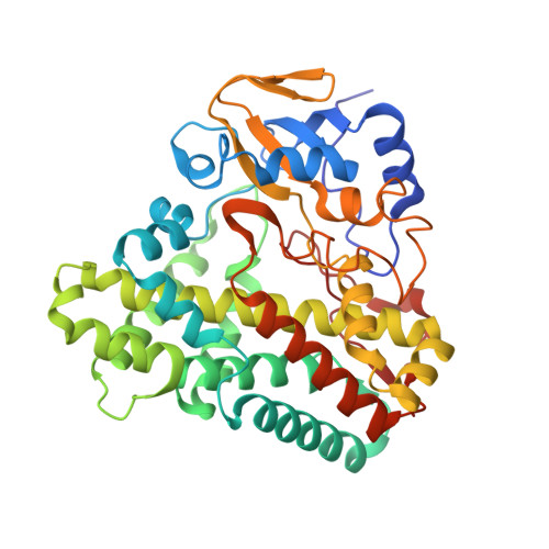
    The essential enzyme CYP121 is a target for drug development against antibiotic resistant strains of Mycobacterium tuberculosis. A triazol-1-yl phenol fragment 1 was identified to bind to CYP121 using a cascade of biophysical assays. Synthetic merging and optimization of 1 produced a 100-fold improvement in binding affinity, yielding lead compound 2 (KD = 15 μM). Deconstruction of 2 into its component retrofragments allowed the group efficiency of structural motifs to be assessed, the identification of more LE scaffolds for optimization and highlighted binding affinity hotspots. Structure-guided addition of a metal-binding pharmacophore onto LE retrofragment scaffolds produced low nanomolar (KD = 15 nM) CYP121 ligands. Elaboration of these compounds to target binding hotspots in the distal active site afforded compounds with excellent selectivity against human drug-metabolizing P450s. Analysis of the factors governing ligand potency and selectivity using X-ray crystallography, UV-vis spectroscopy, and native mass spectrometry provides insight for subsequent drug development.


  • Organizational Affiliation
    • Department of Chemistry, University of Cambridge , Lensfield Road, Cambridge CB2 1EW, U.K.

Macromolecule Content 

  • Total Structure Weight: 44.91 kDa 
  • Atom Count: 3,449 
  • Modeled Residue Count: 393 
  • Deposited Residue Count: 396 
  • Unique protein chains: 1

Macromolecules

Find similar proteins by:|  3D Structure
Entity ID: 1
MoleculeChains  Sequence LengthOrganismDetailsImage
Cytochrome P450 121 CYP121396Mycobacterium tuberculosisMutation(s): 0 
Gene Names: 
EC: 1.14 (PDB Primary Data), 1.14.19.70 (UniProt)
UniProt
Find proteins for P9WPP7 (Mycobacterium tuberculosis (strain ATCC 25618 / H37Rv))
Explore P9WPP7 
Go to UniProtKB:  P9WPP7
Entity Groups
Sequence Clusters30% Identity50% Identity70% Identity90% Identity95% Identity100% Identity
UniProt GroupP9WPP7
Sequence Annotations
Expand
Reference Sequence

Small Molecules

Ligands 3 Unique
IDChains Name / Formula / InChI Key2D Diagram3D Interactions
HEM

Query on HEM


Download:Ideal Coordinates CCD File
E [auth A]PROTOPORPHYRIN IX CONTAINING FE
C34 H32 Fe N4 O4
KABFMIBPWCXCRK-RGGAHWMASA-L
69R

Query on 69R


Download:Ideal Coordinates CCD File
F [auth A],
G [auth A]
4,4'-(5-{[(1H-imidazol-4-yl)methyl]amino}-1H-pyrazole-3,4-diyl)diphenol
C19 H17 N5 O2
QNVXUJBXEJSMQO-UHFFFAOYSA-N
SO4

Query on SO4


Download:Ideal Coordinates CCD File
B [auth A],
C [auth A],
D [auth A]
SULFATE ION
O4 S
QAOWNCQODCNURD-UHFFFAOYSA-L

Experimental Data & Validation

Experimental Data

  • Method: X-RAY DIFFRACTION
  • Resolution: 2.02 Å
  • R-Value Free:  0.203 (Depositor), 0.204 (DCC) 
  • R-Value Work:  0.177 (Depositor), 0.179 (DCC) 
  • R-Value Observed: 0.178 (Depositor) 
Space Group: P 65 2 2
Unit Cell:
Length ( Å )Angle ( ˚ )
a = 77.14α = 90
b = 77.14β = 90
c = 267.27γ = 120
Software Package:
Software NamePurpose
PHENIXrefinement
xia2data reduction
xia2data scaling
PHASERphasing

Structure Validation

View Full Validation Report


Ligand Structure Quality Assessment 


Entry History 

Deposition Data

  • Released Date: 2016-04-06 
  • Deposition Author(s): Levy, C.

Revision History  (Full details and data files)

  • Version 1.0: 2016-04-06
    Type: Initial release
  • Version 1.1: 2016-04-27
    Changes: Database references
  • Version 2.0: 2024-05-01
    Changes: Atomic model, Data collection, Database references, Refinement description