5I9J | pdb_00005i9j

Structure of the cholesterol and lutein-binding domain of human STARD3 at 1.74A


Experimental Data Snapshot

  • Method: X-RAY DIFFRACTION
  • Resolution: 1.74 Å
  • R-Value Free: 
    0.192 (Depositor), 0.198 (DCC) 
  • R-Value Work: 
    0.169 (Depositor), 0.173 (DCC) 
  • R-Value Observed: 
    0.170 (Depositor) 

Starting Model: experimental
View more details

wwPDB Validation 3D Report Full Report

Validation slider image for 5I9J

This is version 1.4 of the entry. See complete history

Literature

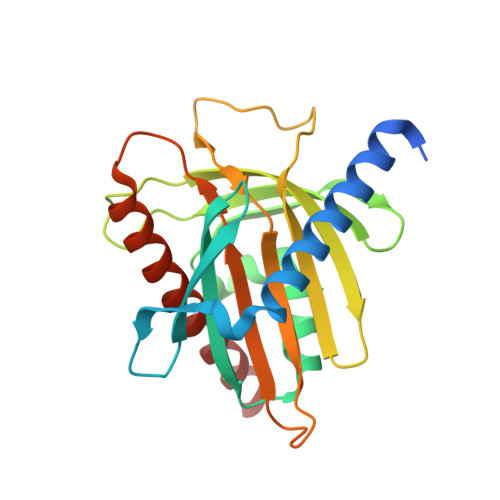
Structure of the lutein-binding domain of human StARD3 at 1.74 angstrom resolution and model of a complex with lutein.

Horvath, M.P.George, E.W.Tran, Q.T.Baumgardner, K.Zharov, G.Lee, S.Sharifzadeh, H.Shihab, S.Mattinson, T.Li, B.Bernstein, P.S.

(2016) Acta Crystallogr F Struct Biol Commun 72: 609-618

  • DOI: https://doi.org/10.1107/S2053230X16010694
  • Primary Citation Related Structures: 
    5I9J

  • PubMed Abstract: 

    A crystal structure of the lutein-binding domain of human StARD3 (StAR-related lipid-transfer protein 3; also known as MLN64) has been refined to 1.74 Å resolution. A previous structure of the same protein determined to 2.2 Å resolution highlighted homology with StARD1 and shared cholesterol-binding character. StARD3 has since been recognized as a carotenoid-binding protein in the primate retina, where its biochemical function of binding lutein with specificity appears to be well suited to recruit this photoprotective molecule. The current and previous structures correspond closely to each other (r.m.s.d. of 0.25 Å), especially in terms of the helix-grip fold constructed around a solvent-filled cavity. Regions of interest were defined with alternate conformations in the current higher-resolution structure, including Arg351 found within the cavity and Ω1, a loop of four residues found just outside the cavity entrance. Models of the complex with lutein generated by rigid-body docking indicate that one of the ionone rings must protrude outside the cavity, and this insight has implications for molecular interactions with transport proteins and enzymes that act on lutein. Interestingly, models with the ℇ-ionone ring characteristic of lutein pointing towards the bottom of the cavity were associated with fewer steric clashes, suggesting that steric complementarity and ligand asymmetry may play a role in discriminating lutein from the other ocular carotenoids zeaxanthin and meso-zeaxanthin, which only have β-ionone rings.


  • Organizational Affiliation
    • Department of Biology, University of Utah, 257 S 1400 E, Salt Lake City, UT 84112, USA.

Macromolecule Content 

  • Total Structure Weight: 26.42 kDa 
  • Atom Count: 2,050 
  • Modeled Residue Count: 214 
  • Deposited Residue Count: 229 
  • Unique protein chains: 1

Macromolecules

Find similar proteins by:|  3D Structure
Entity ID: 1
MoleculeChains  Sequence LengthOrganismDetailsImage
StAR-related lipid transfer protein 3229Homo sapiensMutation(s): 0 
Gene Names: STARD3CAB1MLN64
UniProt & NIH Common Fund Data Resources
Find proteins for Q14849 (Homo sapiens)
Explore Q14849 
Go to UniProtKB:  Q14849
PHAROS:  Q14849
GTEx:  ENSG00000131748 
Entity Groups
Sequence Clusters30% Identity50% Identity70% Identity90% Identity95% Identity100% Identity
UniProt GroupQ14849
Sequence Annotations
Expand
Reference Sequence

Experimental Data & Validation

Experimental Data

  • Method: X-RAY DIFFRACTION
  • Resolution: 1.74 Å
  • R-Value Free:  0.192 (Depositor), 0.198 (DCC) 
  • R-Value Work:  0.169 (Depositor), 0.173 (DCC) 
  • R-Value Observed: 0.170 (Depositor) 
Space Group: P 31 2 1
Unit Cell:
Length ( Å )Angle ( ˚ )
a = 83.39α = 90
b = 83.39β = 90
c = 82.19γ = 120
Software Package:
Software NamePurpose
PHENIXrefinement
XDSdata reduction
XSCALEdata scaling
Cootmodel building
PHASERphasing

Structure Validation

View Full Validation Report


Entry History 

& Funding Information

Deposition Data


Funding OrganizationLocationGrant Number
National Institutes of Health/National Eye Institute (NIH/NEI)United StatesEY11600

Revision History  (Full details and data files)

  • Version 1.0: 2016-03-16
    Type: Initial release
  • Version 1.1: 2016-08-17
    Changes: Database references
  • Version 1.2: 2017-09-27
    Changes: Author supporting evidence, Derived calculations, Refinement description
  • Version 1.3: 2019-12-11
    Changes: Author supporting evidence
  • Version 1.4: 2023-09-27
    Changes: Data collection, Database references, Refinement description