5I8P | pdb_00005i8p

Crystal structure of LP_PLA2 in complex with novel inhibitor


Experimental Data Snapshot

  • Method: X-RAY DIFFRACTION
  • Resolution: 2.37 Å
  • R-Value Free: 
    0.275 (Depositor), 0.273 (DCC) 
  • R-Value Work: 
    0.206 (Depositor), 0.216 (DCC) 
  • R-Value Observed: 
    0.208 (Depositor) 

Starting Model: experimental
View more details

wwPDB Validation 3D Report Full Report

Validation slider image for 5I8P

Ligand Structure Quality Assessment 


This is version 1.2 of the entry. See complete history

Literature

Structural and Thermodynamic Characterization of Protein-Ligand Interactions Formed between Lipoprotein-Associated Phospholipase A2 and Inhibitors

Liu, Q.F.Chen, X.D.Chen, W.Y.Yuan, X.J.Su, H.X.Shen, J.H.Xu, Y.C.

(2016) J Med Chem 59: 5115-5120

  • DOI: https://doi.org/10.1021/acs.jmedchem.6b00282
  • Primary Citation Related Structures: 
    5I8P, 5I9I

  • PubMed Abstract: 

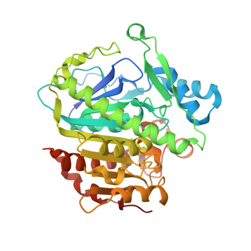
    Lipoprotein-associated phospholipase A2 (Lp-PLA2) represents a promising therapeutic target for atherosclerosis and Alzheimer's disease. Here we reported the first crystal structures of Lp-PLA2 bound with reversible inhibitors and the thermodynamic characterization of complexes. High rigidity of Lp-PLA2 structure and similar binding modes of inhibitors with completely different scaffolds are revealed. It not only provides the molecular basis for inhibitory activity but also sheds light on the essential features of Lp-PLA2 recognition with reversible inhibitors.


  • Organizational Affiliation
    • School of Life Science and Technology, ShanghaiTech University , Shanghai 200031, China.

Macromolecule Content 

  • Total Structure Weight: 88.97 kDa 
  • Atom Count: 5,736 
  • Modeled Residue Count: 735 
  • Deposited Residue Count: 776 
  • Unique protein chains: 1

Macromolecules

Find similar proteins by:|  3D Structure
Entity ID: 1
MoleculeChains  Sequence LengthOrganismDetailsImage
Platelet-activating factor acetylhydrolase
A, B
388Homo sapiensMutation(s): 0 
Gene Names: PLA2G7PAFAH
EC: 3.1.1.47
UniProt & NIH Common Fund Data Resources
Find proteins for Q13093 (Homo sapiens)
Explore Q13093 
Go to UniProtKB:  Q13093
PHAROS:  Q13093
GTEx:  ENSG00000146070 
Entity Groups
Sequence Clusters30% Identity50% Identity70% Identity90% Identity95% Identity100% Identity
UniProt GroupQ13093
Sequence Annotations
Expand
Reference Sequence

Small Molecules

Ligands 1 Unique
IDChains Name / Formula / InChI Key2D Diagram3D Interactions
VQ7

Query on VQ7


Download:Ideal Coordinates CCD File
C [auth A],
D [auth B]
6-[1,1-bis(oxidanylidene)-1,4-thiazinan-4-yl]-4-[[4-[4-chloranyl-3-(trifluoromethyl)phenoxy]-3,5-bis(fluoranyl)phenyl]methoxy]-1-methyl-pyrimidin-2-one
C23 H19 Cl F5 N3 O5 S
FLCSHCSBIFPQJJ-UHFFFAOYSA-N
Binding Affinity Annotations 
IDSourceBinding Affinity
VQ7 BindingDB:  5I8P Kd: 185 (nM) from 1 assay(s)
IC50: 1.7 (nM) from 1 assay(s)

Experimental Data & Validation

Experimental Data

  • Method: X-RAY DIFFRACTION
  • Resolution: 2.37 Å
  • R-Value Free:  0.275 (Depositor), 0.273 (DCC) 
  • R-Value Work:  0.206 (Depositor), 0.216 (DCC) 
  • R-Value Observed: 0.208 (Depositor) 
Space Group: C 1 2 1
Unit Cell:
Length ( Å )Angle ( ˚ )
a = 115.82α = 90
b = 82.75β = 115.44
c = 96.39γ = 90
Software Package:
Software NamePurpose
XSCALEdata scaling
PHASERmodel building
PHENIXrefinement
PDB_EXTRACTdata extraction

Structure Validation

View Full Validation Report


Ligand Structure Quality Assessment 


Entry History 

Deposition Data

Revision History  (Full details and data files)

  • Version 1.0: 2016-06-15
    Type: Initial release
  • Version 1.1: 2017-10-04
    Changes: Data collection, Derived calculations
  • Version 1.2: 2023-11-08
    Changes: Data collection, Database references, Refinement description