5I5H | pdb_00005i5h

Ecoli global domain 245-586


Experimental Data Snapshot

  • Method: X-RAY DIFFRACTION
  • Resolution: 1.65 Å
  • R-Value Free: 
    0.237 (Depositor), 0.231 (DCC) 
  • R-Value Work: 
    0.175 (Depositor), 0.174 (DCC) 
  • R-Value Observed: 
    0.178 (Depositor) 

Starting Model: experimental
View more details

wwPDB Validation 3D Report Full Report

Validation slider image for 5I5H

This is version 2.0 of the entry. See complete history

Literature

Structural insights into cardiolipin transfer from the Inner membrane to the outer membrane by PbgA in Gram-negative bacteria.

Dong, H.Zhang, Z.Tang, X.Huang, S.Li, H.Peng, B.Dong, C.

(2016) Sci Rep 6: 30815-30815

  • DOI: https://doi.org/10.1038/srep30815
  • Primary Citation Related Structures: 
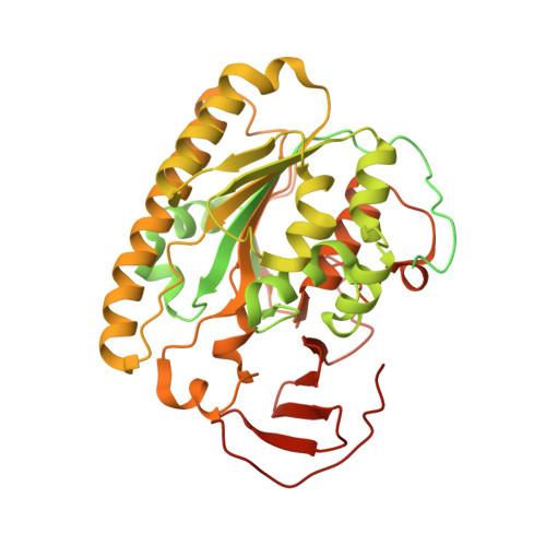
    5I5D, 5I5F, 5I5H

  • PubMed Abstract: 

    The outer membrane (OM) of Gram-negative bacteria is a unique asymmetric lipid bilayer in which the outer leaflet is composed of lipopolysaccharide (LPS) and the inner leaflet is formed by glycerophospholipid (GPL). The OM plays a fundamental role in protecting Gram-negative bacteria from harsh environments and toxic compounds. The transport and assembly pathways for phospholipids of bacterial OM are unknown. Cardiolipin (CL) plays an important role in OM biogenesis and pathogenesis, and the inner membrane (IM) protein PbgA, containing five transmembrane domains and a globular domain in periplasm has been recently identified as a CL transporter from the IM to the OM with an unknown mechanism. Here we present the first two crystal structures of soluble periplasmic globular domain of PbgA from S. typhimurium and E. coli, which revealed that the globular domains of PbgA resemble the structures of the arylsulfatase protein family and contains a novel core hydrophobic pocket that may be responsible for binding and transporting CLs. Our structural and functional studies shed an important light on the mechanism of CL transport in Gram-negative bacteria from the IM to the OM, which offers great potential for the development of novel antibiotics against multi-drug resistant bacterial infections.


  • Organizational Affiliation
    • Biomedical Research Centre, Norwich Medical School, University of East Anglia, Norwich Research Park, Norwich, NR4 7TJ, UK.

Macromolecule Content 

  • Total Structure Weight: 67.36 kDa 
  • Atom Count: 3,099 
  • Modeled Residue Count: 327 
  • Deposited Residue Count: 586 
  • Unique protein chains: 1

Macromolecules

Find similar proteins by:|  3D Structure
Entity ID: 1
MoleculeChains  Sequence LengthOrganismDetailsImage
Inner membrane protein YejM586Escherichia coli K-12Mutation(s): 0 
Gene Names: yejMyejNb2188JW2176
EC: 3.1
Membrane Entity: Yes 
UniProt
Find proteins for P0AD27 (Escherichia coli (strain K12))
Explore P0AD27 
Go to UniProtKB:  P0AD27
Entity Groups
Sequence Clusters30% Identity50% Identity70% Identity90% Identity95% Identity100% Identity
UniProt GroupP0AD27
Sequence Annotations
Expand
Reference Sequence

Experimental Data & Validation

Experimental Data

  • Method: X-RAY DIFFRACTION
  • Resolution: 1.65 Å
  • R-Value Free:  0.237 (Depositor), 0.231 (DCC) 
  • R-Value Work:  0.175 (Depositor), 0.174 (DCC) 
  • R-Value Observed: 0.178 (Depositor) 
Space Group: P 31 2 1
Unit Cell:
Length ( Å )Angle ( ˚ )
a = 56.86α = 90
b = 56.86β = 90
c = 191.81γ = 120
Software Package:
Software NamePurpose
REFMACrefinement
XDSdata reduction
XDSdata scaling
PHASERphasing

Structure Validation

View Full Validation Report


Entry History 

Deposition Data

Revision History  (Full details and data files)

  • Version 1.0: 2016-08-17
    Type: Initial release
  • Version 2.0: 2024-01-10
    Changes: Atomic model, Data collection, Database references, Refinement description