5I2S

PREFUSION FORM OF THE VESICULAR STOMATITIS VIRUS GLYCOPROTEIN G ECTODOMAIN


Experimental Data Snapshot

  • Method: X-RAY DIFFRACTION
  • Resolution: 3.00 Å
  • R-Value Free: 0.239 
  • R-Value Work: 0.181 
  • R-Value Observed: 0.184 

wwPDB Validation   3D Report Full Report


Ligand Structure Quality Assessment 


This is version 2.1 of the entry. See complete history


Literature

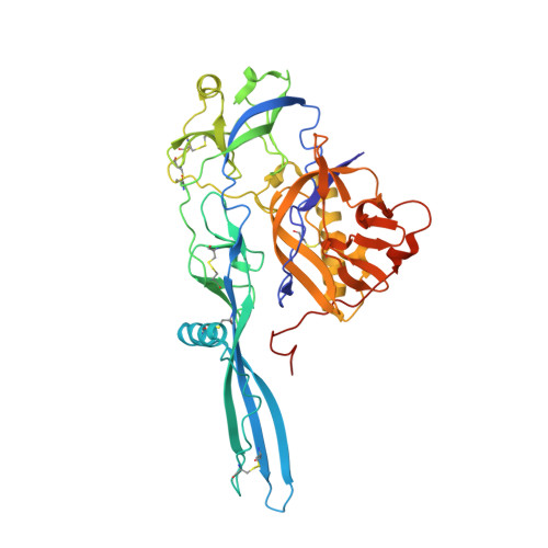
Structure of the prefusion form of the vesicular stomatitis virus glycoprotein G.

Roche, S.Rey, F.A.Gaudin, Y.Bressanelli, S.

(2007) Science 315: 843-848

  • DOI: https://doi.org/10.1126/science.1135710
  • Primary Citation of Related Structures:  
    5I2S

  • PubMed Abstract: 

    Glycoprotein G of the vesicular stomatitis virus triggers membrane fusion via a low pH-induced structural rearrangement. Despite the equilibrium between the pre- and postfusion states, the structure of the prefusion form, determined to 3.0 angstrom resolution, shows that the fusogenic transition entails an extensive structural reorganization of G. Comparison with the structure of the postfusion form suggests a pathway for the conformational change. In the prefusion form, G has the shape of a tripod with the fusion loops exposed, which point toward the viral membrane, and with the antigenic sites located at the distal end of the molecule. A large number of G glycoproteins, perhaps organized as in the crystals, act cooperatively to induce membrane merging.


  • Organizational Affiliation

    CNRS, Unité Mixte de Recherche (UMR) 2472, Institut National de la Recherche Agronomique (INRA), UMR 1157, Institut Fédératif de Recherche 115, Laboratoire de Virologie Moléculaire et Structurale, 91198, Gif sur Yvette, France.


Macromolecules
Find similar proteins by:  (by identity cutoff)  |  3D Structure
Entity ID: 1
MoleculeChains Sequence LengthOrganismDetailsImage
Glycoprotein G422Vesicular stomatitis Indiana virus strain Mudd-SummersMutation(s): 0 
Membrane Entity: Yes 
UniProt
Find proteins for P0C2X0 (Vesicular stomatitis Indiana virus (strain Mudd-Summers))
Explore P0C2X0 
Go to UniProtKB:  P0C2X0
Entity Groups  
Sequence Clusters30% Identity50% Identity70% Identity90% Identity95% Identity100% Identity
UniProt GroupP0C2X0
Sequence Annotations
Expand
  • Reference Sequence
Oligosaccharides

Help

Entity ID: 2
MoleculeChains Length2D Diagram Glycosylation3D Interactions
2-acetamido-2-deoxy-beta-D-glucopyranose-(1-4)-2-acetamido-2-deoxy-beta-D-glucopyranose
B
2N-Glycosylation
Glycosylation Resources
GlyTouCan:  G42666HT
GlyCosmos:  G42666HT
GlyGen:  G42666HT
Experimental Data & Validation

Experimental Data

  • Method: X-RAY DIFFRACTION
  • Resolution: 3.00 Å
  • R-Value Free: 0.239 
  • R-Value Work: 0.181 
  • R-Value Observed: 0.184 
  • Space Group: P 6 2 2
Unit Cell:
Length ( Å )Angle ( ˚ )
a = 120.51α = 90
b = 120.51β = 90
c = 183.13γ = 120
Software Package:
Software NamePurpose
PHENIXrefinement
XDSdata reduction
XSCALEdata scaling
PHASERphasing

Structure Validation

View Full Validation Report


Ligand Structure Quality Assessment 


Entry History 

Deposition Data

Revision History  (Full details and data files)

  • Version 1.0: 2016-03-02
    Type: Initial release
  • Version 2.0: 2020-07-29
    Type: Remediation
    Reason: Carbohydrate remediation
    Changes: Atomic model, Data collection, Derived calculations, Structure summary
  • Version 2.1: 2024-01-10
    Changes: Data collection, Database references, Refinement description, Structure summary