5I2A | pdb_00005i2a

1,2-propanediol Dehydration in Roseburia inulinivorans; Structural Basis for Substrate and Enantiomer Selectivity


Experimental Data Snapshot

  • Method: X-RAY DIFFRACTION
  • Resolution: 2.10 Å
  • R-Value Free: 
    0.183 (Depositor), 0.186 (DCC) 
  • R-Value Work: 
    0.152 (Depositor), 0.159 (DCC) 
  • R-Value Observed: 
    0.153 (Depositor) 

wwPDB Validation 3D Report Full Report

Validation slider image for 5I2A

This is version 1.3 of the entry. See complete history

Literature

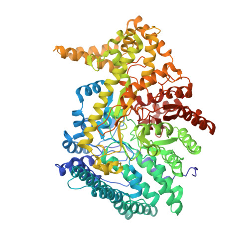
1,2-Propanediol Dehydration in Roseburia inulinivorans: STRUCTURAL BASIS FOR SUBSTRATE AND ENANTIOMER SELECTIVITY.

LaMattina, J.W.Keul, N.D.Reitzer, P.Kapoor, S.Galzerani, F.Koch, D.J.Gouvea, I.E.Lanzilotta, W.N.

(2016) J Biological Chem 291: 15515-15526

  • DOI: https://doi.org/10.1074/jbc.M116.721142
  • Primary Citation Related Structures: 
    5I2A, 5I2G

  • PubMed Abstract: 

    Glycyl radical enzymes (GREs) represent a diverse superfamily of enzymes that utilize a radical mechanism to catalyze difficult, but often essential, chemical reactions. In this work we present the first biochemical and structural data for a GRE-type diol dehydratase from the organism Roseburia inulinivorans (RiDD). Despite high sequence (48% identity) and structural similarity to the GRE-type glycerol dehydratase from Clostridium butyricum, we demonstrate that the RiDD is in fact a diol dehydratase. In addition, the RiDD will utilize both (S)-1,2-propanediol and (R)-1,2-propanediol as a substrate, with an observed preference for the S enantiomer. Based on the new structural information we developed and successfully tested a hypothesis that explains the functional differences we observe.


  • Organizational Affiliation
    • From the Department of Biochemistry and Molecular Biology, University of Georgia, Athens, Georgia 30602 and.

Macromolecule Content 

  • Total Structure Weight: 188.88 kDa 
  • Atom Count: 14,399 
  • Modeled Residue Count: 1,673 
  • Deposited Residue Count: 1,686 
  • Unique protein chains: 1

Macromolecules

Find similar proteins by:|  3D Structure
Entity ID: 1
MoleculeChains  Sequence LengthOrganismDetailsImage
Diol-dehydratase
A, B
843Roseburia inulinivoransMutation(s): 0 
UniProt
Find proteins for Q1A666 (Roseburia inulinivorans)
Explore Q1A666 
Go to UniProtKB:  Q1A666
Entity Groups
Sequence Clusters30% Identity50% Identity70% Identity90% Identity95% Identity100% Identity
UniProt GroupQ1A666
Sequence Annotations
Expand
Reference Sequence

Experimental Data & Validation

Experimental Data

  • Method: X-RAY DIFFRACTION
  • Resolution: 2.10 Å
  • R-Value Free:  0.183 (Depositor), 0.186 (DCC) 
  • R-Value Work:  0.152 (Depositor), 0.159 (DCC) 
  • R-Value Observed: 0.153 (Depositor) 
Space Group: C 2 2 21
Unit Cell:
Length ( Å )Angle ( ˚ )
a = 138.206α = 90
b = 183.225β = 90
c = 228.339γ = 90
Software Package:
Software NamePurpose
PHENIXrefinement
PDB_EXTRACTdata extraction
HKL-2000data reduction
SCALEPACKdata scaling
MOLREPphasing

Structure Validation

View Full Validation Report


Entry History 

Deposition Data

Revision History  (Full details and data files)

  • Version 1.0: 2016-06-01
    Type: Initial release
  • Version 1.1: 2016-06-15
    Changes: Database references, Structure summary
  • Version 1.2: 2016-08-10
    Changes: Database references
  • Version 1.3: 2024-03-06
    Changes: Data collection, Database references, Derived calculations