5HZH | pdb_00005hzh

Crystal structure of photoinhibitable Rac1 containing C450A mutant LOV2 domain


Experimental Data Snapshot

  • Method: X-RAY DIFFRACTION
  • Resolution: 2.60 Å
  • R-Value Free: 
    0.255 (Depositor), 0.260 (DCC) 
  • R-Value Work: 
    0.204 (Depositor), 0.206 (DCC) 
  • R-Value Observed: 
    0.206 (Depositor) 

Starting Model: experimental
View more details

wwPDB Validation 3D Report Full Report

Validation slider image for 5HZH

Ligand Structure Quality Assessment 


This is version 1.2 of the entry. See complete history

Literature

Engineering extrinsic disorder to control protein activity in living cells.

Dagliyan, O.Tarnawski, M.Chu, P.H.Shirvanyants, D.Schlichting, I.Dokholyan, N.V.Hahn, K.M.

(2016) Science 354: 1441-1444

  • DOI: https://doi.org/10.1126/science.aah3404
  • Primary Citation Related Structures: 
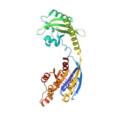
    5HZH, 5HZI, 5HZJ, 5HZK

  • PubMed Abstract: 

    Optogenetic and chemogenetic control of proteins has revealed otherwise inaccessible facets of signaling dynamics. Here, we use light- or ligand-sensitive domains to modulate the structural disorder of diverse proteins, thereby generating robust allosteric switches. Sensory domains were inserted into nonconserved, surface-exposed loops that were tight and identified computationally as allosterically coupled to active sites. Allosteric switches introduced into motility signaling proteins (kinases, guanosine triphosphatases, and guanine exchange factors) controlled conversion between conformations closely resembling natural active and inactive states, as well as modulated the morphodynamics of living cells. Our results illustrate a broadly applicable approach to design physiological protein switches.


  • Organizational Affiliation
    • Program in Molecular and Cellular Biophysics, University of North Carolina at Chapel Hill, Chapel Hill, NC 27599, USA.

Macromolecule Content 

  • Total Structure Weight: 38.15 kDa 
  • Atom Count: 2,622 
  • Modeled Residue Count: 320 
  • Deposited Residue Count: 332 
  • Unique protein chains: 1

Macromolecules

Find similar proteins by:|  3D Structure
Entity ID: 1
MoleculeChains  Sequence LengthOrganismDetailsImage
Ras-related C3 botulinum toxin substrate 1,NPH1-1,Ras-related C3 botulinum toxin substrate 1332Homo sapiensAvena sativaMutation(s): 2 
Gene Names: RAC1TC25MIG5NPH1-1
EC: 2.7.11.1 (UniProt), 3.6.5.2 (UniProt)
UniProt & NIH Common Fund Data Resources
Find proteins for O49003 (Avena sativa)
Explore O49003 
Go to UniProtKB:  O49003
Find proteins for P63000 (Homo sapiens)
Explore P63000 
Go to UniProtKB:  P63000
PHAROS:  P63000
GTEx:  ENSG00000136238 
Entity Groups
Sequence Clusters30% Identity50% Identity70% Identity90% Identity95% Identity100% Identity
UniProt GroupsP63000O49003
Sequence Annotations
Expand
Reference Sequence

Experimental Data & Validation

Experimental Data

  • Method: X-RAY DIFFRACTION
  • Resolution: 2.60 Å
  • R-Value Free:  0.255 (Depositor), 0.260 (DCC) 
  • R-Value Work:  0.204 (Depositor), 0.206 (DCC) 
  • R-Value Observed: 0.206 (Depositor) 
Space Group: P 1 21 1
Unit Cell:
Length ( Å )Angle ( ˚ )
a = 36.05α = 90
b = 74.27β = 94.62
c = 58.4γ = 90
Software Package:
Software NamePurpose
PHENIXrefinement
XDSdata reduction
XSCALEdata scaling
PHASERphasing

Structure Validation

View Full Validation Report


Ligand Structure Quality Assessment 


Entry History 

Deposition Data

Revision History  (Full details and data files)

  • Version 1.0: 2016-12-21
    Type: Initial release
  • Version 1.1: 2016-12-28
    Changes: Database references
  • Version 1.2: 2024-01-10
    Changes: Data collection, Database references, Derived calculations, Refinement description