5HWZ | pdb_00005hwz

Crystal structure of nitrophorin 4 D30N mutant with nitrite


Experimental Data Snapshot

  • Method: X-RAY DIFFRACTION
  • Resolution: 1.45 Å
  • R-Value Free: 
    0.180 (Depositor), 0.178 (DCC) 
  • R-Value Work: 
    0.149 (Depositor), 0.145 (DCC) 
  • R-Value Observed: 
    0.150 (Depositor) 

Starting Model: experimental
View more details

wwPDB Validation 3D Report Full Report

Validation slider image for 5HWZ

Ligand Structure Quality Assessment 


This is version 2.2 of the entry. See complete history

Literature

Elucidation of the heme active site electronic structure affecting the unprecedented nitrite dismutase activity of the ferrihemebproteins, the nitrophorins.

He, C.Ogata, H.Lubitz, W.

(2016) Chem Sci 7: 5332-5340

  • DOI: https://doi.org/10.1039/c6sc01019a
  • Primary Citation Related Structures: 
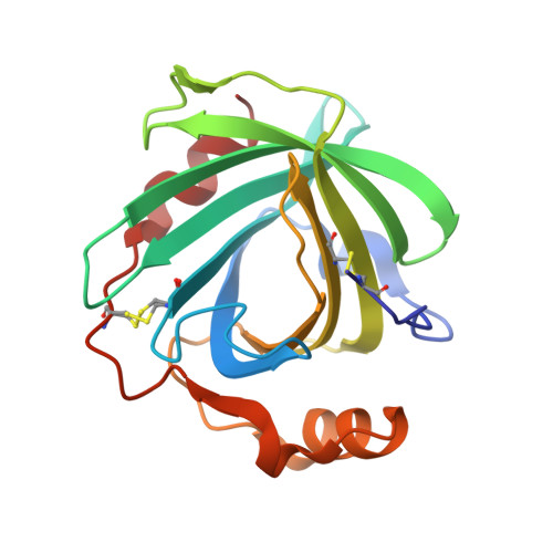
    5HWZ

  • PubMed Abstract: 

    Nitrophorins (NPs) catalyze the nitrite dismutation reaction that is unprecedented in ferriheme proteins. Despite progress in studying the reaction mechanism, fundamental issues regarding the correlation of the structural features with the nitrite dismutase activity of NPs remain elusive. On the other hand, it has been shown that the nitrite complexes of NPs are unique among those of the ferriheme proteins since some of their electron paramagnetic resonance (EPR) spectra show significant highly anisotropic low spin (HALS) signals with large g max values over 3.2. The origin of HALS signals in ferriheme proteins or models is not well understood, especially in cases where axial ligands other than histidine are present. In this study several mutations were introduced in NP4. The related nitrite coordination and dismutation reaction were investigated. As a result, the EPR spectra of the NP-nitrite complexes were found to be tightly correlated with the extent of heme ruffling and protonation state of the proximal His ligand-dictated by an extended H-bonding network at the heme active site. Furthermore, it is established that the two factors are essential in determining the nitrite dismutase activity of NPs. These results may provide a valuable guide for identifying or designing novel heme proteins with similar activity.


  • Organizational Affiliation
    • Max Planck Institute for Chemical Energy Conversion , Stiftstrasse 34-36 , D-45470 , Mülheim an der Ruhr , Germany . Email: chunmao82@gmail.com ; Email: wolfgang.lubitz@cec.mpg.de.

Macromolecule Content 

  • Total Structure Weight: 20.95 kDa 
  • Atom Count: 1,662 
  • Modeled Residue Count: 184 
  • Deposited Residue Count: 184 
  • Unique protein chains: 1

Macromolecules

Find similar proteins by:|  3D Structure
Entity ID: 1
MoleculeChains  Sequence LengthOrganismDetailsImage
Nitrophorin-4184Rhodnius prolixusMutation(s): 1 
EC: 1.7.6.1
UniProt
Find proteins for Q94734 (Rhodnius prolixus)
Explore Q94734 
Go to UniProtKB:  Q94734
Entity Groups
Sequence Clusters30% Identity50% Identity70% Identity90% Identity95% Identity100% Identity
UniProt GroupQ94734
Sequence Annotations
Expand
Reference Sequence

Small Molecules

Experimental Data & Validation

Experimental Data

  • Method: X-RAY DIFFRACTION
  • Resolution: 1.45 Å
  • R-Value Free:  0.180 (Depositor), 0.178 (DCC) 
  • R-Value Work:  0.149 (Depositor), 0.145 (DCC) 
  • R-Value Observed: 0.150 (Depositor) 
Space Group: C 1 2 1
Unit Cell:
Length ( Å )Angle ( ˚ )
a = 69.779α = 90
b = 43.11β = 94.54
c = 52.623γ = 90
Software Package:
Software NamePurpose
PHENIXrefinement
XDSdata reduction
XSCALEdata scaling
MOLREPphasing

Structure Validation

View Full Validation Report


Ligand Structure Quality Assessment 


Entry History 

Deposition Data

Revision History  (Full details and data files)

  • Version 1.0: 2016-05-04
    Type: Initial release
  • Version 2.0: 2018-09-12
    Changes: Atomic model, Data collection, Database references
  • Version 2.1: 2024-01-10
    Changes: Data collection, Database references, Refinement description
  • Version 2.2: 2024-11-06
    Changes: Structure summary