5HW0 | pdb_00005hw0

Erwinia chrysanthemi L-asparaginase + Glutamic acid

  • Classification: HYDROLASE
  • Organism(s): Dickeya chrysanthemi
  • Expression System: Escherichia coli
  • Mutation(s): No 

  • Deposited: 2016-01-28 Released: 2016-04-06 
  • Deposition Author(s): Nguyen, H.A., Lavie, A.
  • Funding Organization(s): National Institutes of Health/National Institute of Biomedical Imaging and Bioengineering (NIH/NIBIB)

Experimental Data Snapshot

  • Method: X-RAY DIFFRACTION
  • Resolution: 1.70 Å
  • R-Value Free: 
    0.205 (Depositor), 0.206 (DCC) 
  • R-Value Work: 
    0.167 (Depositor), 0.167 (DCC) 
  • R-Value Observed: 
    0.168 (Depositor) 

Starting Model: experimental
View more details

wwPDB Validation 3D Report Full Report

Validation slider image for 5HW0

This is version 1.4 of the entry. See complete history

Literature

Structural Insight into Substrate Selectivity of Erwinia chrysanthemi l-Asparaginase.

Nguyen, H.A.Su, Y.Lavie, A.

(2016) Biochemistry 55: 1246-1253

  • DOI: https://doi.org/10.1021/acs.biochem.5b01351
  • Primary Citation Related Structures: 
    5F52, 5HW0

  • PubMed Abstract: 

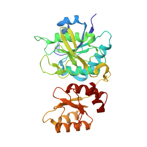
    l-Asparaginases of bacterial origin are a mainstay of acute lymphoblastic leukemia treatment. The mechanism of action of these enzyme drugs is associated with their capacity to deplete the amino acid l-asparagine from the blood. However, clinical use of bacterial l-asparaginases is complicated by their dual l-asparaginase and l-glutaminase activities. The latter, even though representing only ∼10% of the overall activity, is partially responsible for the observed toxic side effects. Hence, l-asparaginases devoid of l-glutaminase activity hold potential as safer drugs. Understanding the key determinants of l-asparaginase substrate specificity is a prerequisite step toward the development of enzyme variants with reduced toxicity. Here we present crystal structures of the Erwinia chrysanthemi l-asparaginase in complex with l-aspartic acid and with l-glutamic acid. These structures reveal two enzyme conformations-open and closed-corresponding to the inactive and active states, respectively. The binding of ligands induces the positioning of the catalytic Thr15 into its active conformation, which in turn allows for the ordering and closure of the flexible N-terminal loop. Notably, l-aspartic acid is more efficient than l-glutamic acid in inducing the active positioning of Thr15. Structural elements explaining the preference of the enzyme for l-asparagine over l-glutamine are discussed with guidance to the future development of more specific l-asparaginases.


  • Organizational Affiliation
    • The Jesse Brown VA Medical Center , Chicago, Illinois 60607, United States.

Macromolecule Content 

  • Total Structure Weight: 141.76 kDa 
  • Atom Count: 10,718 
  • Modeled Residue Count: 1,255 
  • Deposited Residue Count: 1,312 
  • Unique protein chains: 1

Macromolecules

Find similar proteins by:|  3D Structure
Entity ID: 1
MoleculeChains  Sequence LengthOrganismDetailsImage
L-asparaginase
A, B, C, D
328Dickeya chrysanthemiMutation(s): 0 
Gene Names: ansBasn
EC: 3.5.1.1
UniProt
Find proteins for P06608 (Dickeya chrysanthemi)
Explore P06608 
Go to UniProtKB:  P06608
Entity Groups
Sequence Clusters30% Identity50% Identity70% Identity90% Identity95% Identity100% Identity
UniProt GroupP06608
Sequence Annotations
Expand
Reference Sequence

Experimental Data & Validation

Experimental Data

  • Method: X-RAY DIFFRACTION
  • Resolution: 1.70 Å
  • R-Value Free:  0.205 (Depositor), 0.206 (DCC) 
  • R-Value Work:  0.167 (Depositor), 0.167 (DCC) 
  • R-Value Observed: 0.168 (Depositor) 
Space Group: P 21 21 21
Unit Cell:
Length ( Å )Angle ( ˚ )
a = 75.24α = 90
b = 88.818β = 90
c = 174.573γ = 90
Software Package:
Software NamePurpose
PHENIXrefinement
XDSdata scaling
XDSdata reduction
PDB_EXTRACTdata extraction
MOLREPphasing

Structure Validation

View Full Validation Report


Entry History 

& Funding Information

Deposition Data


Funding OrganizationLocationGrant Number
National Institutes of Health/National Institute of Biomedical Imaging and Bioengineering (NIH/NIBIB)United StatesRO1 EB013685

Revision History  (Full details and data files)

  • Version 1.0: 2016-04-06
    Type: Initial release
  • Version 1.1: 2016-07-06
    Changes: Data collection
  • Version 1.2: 2017-09-27
    Changes: Advisory, Author supporting evidence, Derived calculations
  • Version 1.3: 2019-12-11
    Changes: Author supporting evidence
  • Version 1.4: 2023-09-27
    Changes: Advisory, Data collection, Database references, Refinement description