5HVS | pdb_00005hvs

Crystal Structure of Macrophage Migration Inhibitory Factor (MIF) with a Biaryltriazole Inhibitor (3i-305)


Experimental Data Snapshot

  • Method: X-RAY DIFFRACTION
  • Resolution: 1.75 Å
  • R-Value Free: 
    0.206 (Depositor), 0.211 (DCC) 
  • R-Value Work: 
    0.170 (Depositor), 0.181 (DCC) 
  • R-Value Observed: 
    0.174 (Depositor) 

Starting Model: experimental
View more details

wwPDB Validation 3D Report Full Report

Validation slider image for 5HVS

Ligand Structure Quality Assessment 


This is version 1.4 of the entry. See complete history

Literature

A Fluorescence Polarization Assay for Binding to Macrophage Migration Inhibitory Factor and Crystal Structures for Complexes of Two Potent Inhibitors.

Cisneros, J.A.Robertson, M.J.Valhondo, M.Jorgensen, W.L.

(2016) J Am Chem Soc 138: 8630-8638

  • DOI: https://doi.org/10.1021/jacs.6b04910
  • Primary Citation Related Structures: 
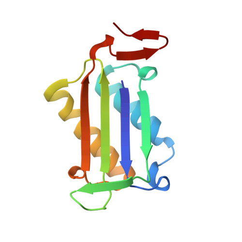
    5HVS, 5HVT

  • PubMed Abstract: 

    Human macrophage migration inhibitory factor (MIF) is both a keto-enol tautomerase and a cytokine associated with numerous inflammatory diseases and cancer. Consistent with observed correlations between inhibition of the enzymatic and biological activities, discovery of MIF inhibitors has focused on monitoring the tautomerase activity using l-dopachrome methyl ester or 4-hydroxyphenyl pyruvic acid as substrates. The accuracy of these assays is compromised by several issues including substrate instability, spectral interference, and short linear periods for product formation. In this work, we report the syntheses of fluorescently labeled MIF inhibitors and their use in the first fluorescence polarization-based assay to measure the direct binding of inhibitors to the active site. The assay allows the accurate and efficient identification of competitive, noncompetitive, and covalent inhibitors of MIF in a manner that can be scaled for high-throughput screening. The results for 22 compounds show that the most potent MIF inhibitors bind with Kd values of ca. 50 nM; two are from our laboratory, and the other is a compound from the patent literature. X-ray crystal structures for two of the most potent compounds bound to MIF are also reported here. Striking combinations of protein-ligand hydrogen bonding, aryl-aryl, and cation-π interactions are responsible for the high affinities. A new chemical series was then designed using this knowledge to yield two more strong MIF inhibitors/binders.


  • Organizational Affiliation
    • Department of Chemistry, Yale University , New Haven, Connecticut 06520-8107, United States.

Macromolecule Content 

  • Total Structure Weight: 39.15 kDa 
  • Atom Count: 3,035 
  • Modeled Residue Count: 342 
  • Deposited Residue Count: 342 
  • Unique protein chains: 1

Macromolecules

Find similar proteins by:|  3D Structure
Entity ID: 1
MoleculeChains  Sequence LengthOrganismDetailsImage
Macrophage migration inhibitory factor
A, B, C
114Homo sapiensMutation(s): 0 
Gene Names: MIFGLIFMMIF
EC: 5.3.2.1 (PDB Primary Data), 5.3.3.12 (PDB Primary Data)
UniProt & NIH Common Fund Data Resources
Find proteins for P14174 (Homo sapiens)
Explore P14174 
Go to UniProtKB:  P14174
PHAROS:  P14174
GTEx:  ENSG00000240972 
Entity Groups
Sequence Clusters30% Identity50% Identity70% Identity90% Identity95% Identity100% Identity
UniProt GroupP14174
Sequence Annotations
Expand
Reference Sequence

Small Molecules

Ligands 3 Unique
IDChains Name / Formula / InChI Key2D Diagram3D Interactions
65V

Query on 65V


Download:Ideal Coordinates CCD File
F [auth A],
I [auth B],
M [auth C]
3-({2-[1-(3-fluoro-4-hydroxyphenyl)-1H-1,2,3-triazol-4-yl]quinolin-5-yl}oxy)benzoic acid
C24 H15 F N4 O4
RXHQCISHMPQZDT-UHFFFAOYSA-N
SO4

Query on SO4


Download:Ideal Coordinates CCD File
D [auth A]
E [auth A]
G [auth B]
H [auth B]
J [auth B]
D [auth A],
E [auth A],
G [auth B],
H [auth B],
J [auth B],
L [auth C]
SULFATE ION
O4 S
QAOWNCQODCNURD-UHFFFAOYSA-L
GOL

Query on GOL


Download:Ideal Coordinates CCD File
K [auth B],
N [auth C]
GLYCEROL
C3 H8 O3
PEDCQBHIVMGVHV-UHFFFAOYSA-N
Binding Affinity Annotations 
IDSourceBinding Affinity
65V BindingDB:  5HVS Ki: min: 14, max: 57 (nM) from 3 assay(s)

Experimental Data & Validation

Experimental Data

  • Method: X-RAY DIFFRACTION
  • Resolution: 1.75 Å
  • R-Value Free:  0.206 (Depositor), 0.211 (DCC) 
  • R-Value Work:  0.170 (Depositor), 0.181 (DCC) 
  • R-Value Observed: 0.174 (Depositor) 
Space Group: I 2 2 2
Unit Cell:
Length ( Å )Angle ( ˚ )
a = 68.531α = 90
b = 69.744β = 90
c = 133.793γ = 90
Software Package:
Software NamePurpose
REFMACrefinement
HKL-2000data scaling
PDB_EXTRACTdata extraction
HKL-2000data reduction
PHASERphasing

Structure Validation

View Full Validation Report


Ligand Structure Quality Assessment 


Entry History 

& Funding Information

Deposition Data


Funding OrganizationLocationGrant Number
National Institutes of Health/National Institute of General Medical Sciences (NIH/NIGMS)United StatesGM32136

Revision History  (Full details and data files)

  • Version 1.0: 2016-06-29
    Type: Initial release
  • Version 1.1: 2016-07-20
    Changes: Database references
  • Version 1.2: 2017-09-27
    Changes: Author supporting evidence, Derived calculations
  • Version 1.3: 2019-12-25
    Changes: Author supporting evidence
  • Version 1.4: 2023-09-27
    Changes: Data collection, Database references, Refinement description