5HSW | pdb_00005hsw

KSHV SOX RNA complex


Experimental Data Snapshot

  • Method: X-RAY DIFFRACTION
  • Resolution: 3.30 Å
  • R-Value Free: 
    0.262 (Depositor), 0.266 (DCC) 
  • R-Value Work: 
    0.208 (Depositor), 0.221 (DCC) 
  • R-Value Observed: 
    0.211 (Depositor) 

Starting Model: experimental
View more details

wwPDB Validation 3D Report Full Report

Validation slider image for 5HSW

This is version 1.5 of the entry. See complete history

Literature

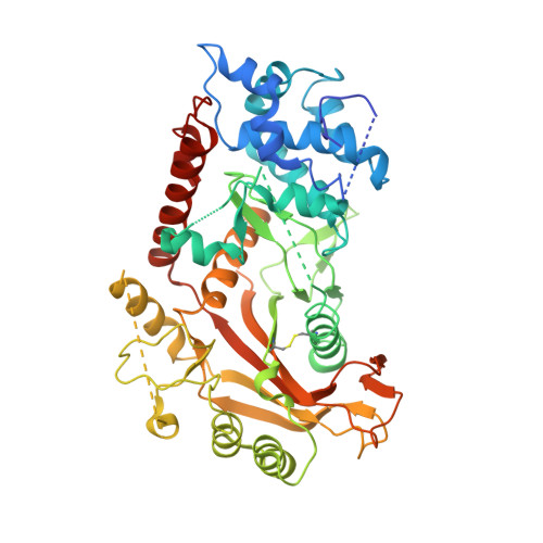
KSHV SOX mediated host shutoff: the molecular mechanism underlying mRNA transcript processing.

Lee, H.Patschull, A.O.M.Bagneris, C.Ryan, H.Sanderson, C.M.Ebrahimi, B.Nobeli, I.Barrett, T.E.

(2017) Nucleic Acids Res 45: 4756-4767

  • DOI: https://doi.org/10.1093/nar/gkw1340
  • Primary Citation Related Structures: 
    5HSW

  • PubMed Abstract: 

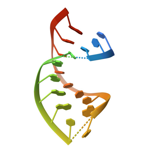
    Onset of the lytic phase in the KSHV life cycle is accompanied by the rapid, global degradation of host (and viral) mRNA transcripts in a process termed host shutoff. Key to this destruction is the virally encoded alkaline exonuclease SOX. While SOX has been shown to possess an intrinsic RNase activity and a potential consensus sequence for endonucleolytic cleavage identified, the structures of the RNA substrates targeted remained unclear. Based on an analysis of three reported target transcripts, we were able to identify common structures and confirm that these are indeed degraded by SOX in vitro as well as predict the presence of such elements in the KSHV pre-microRNA transcript K12-2. From these studies, we were able to determine the crystal structure of SOX productively bound to a 31 nucleotide K12-2 fragment. This complex not only reveals the structural determinants required for RNA recognition and degradation but, together with biochemical and biophysical studies, reveals distinct roles for residues implicated in host shutoff. Our results further confirm that SOX and the host exoribonuclease Xrn1 act in concert to elicit the rapid degradation of mRNA substrates observed in vivo, and that the activities of the two ribonucleases are co-ordinated.


  • Organizational Affiliation
    • Institute for Structural and Molecular Biology, Department of Biological Sciences, Birkbeck College, Malet Street, London WC1E 7HX, UK.

Macromolecule Content 

  • Total Structure Weight: 65.24 kDa 
  • Atom Count: 3,625 
  • Modeled Residue Count: 446 
  • Deposited Residue Count: 519 
  • Unique protein chains: 1
  • Unique nucleic acid chains: 1

Macromolecules


Find similar proteins by:|  3D Structure
Entity ID: 1
MoleculeChains  Sequence LengthOrganismDetailsImage
ORF 37488Human herpesvirus 8 type MMutation(s): 1 
UniProt
Find proteins for P88925 (Human herpesvirus 8 type M)
Explore P88925 
Go to UniProtKB:  P88925
Entity Groups
Sequence Clusters30% Identity50% Identity70% Identity90% Identity95% Identity100% Identity
UniProt GroupP88925
Sequence Annotations
Expand
Reference Sequence
Find similar nucleic acids by:  (by identity cutoff) 
Entity ID: 2
MoleculeChains LengthOrganismImage
RNA (5'-R(P*UP*CP*UP*UP*GP*AP*AP*GP*CP*AP*GP*CP*UP*UP*CP*CP*AP*G)-3')31Human gammaherpesvirus 8
Sequence Annotations
Expand
Reference Sequence

Small Molecules

Ligands 1 Unique
IDChains Name / Formula / InChI Key2D Diagram3D Interactions
ACT

Query on ACT


Download:Ideal Coordinates CCD File
C [auth A]ACETATE ION
C2 H3 O2
QTBSBXVTEAMEQO-UHFFFAOYSA-M

Experimental Data & Validation

Experimental Data

  • Method: X-RAY DIFFRACTION
  • Resolution: 3.30 Å
  • R-Value Free:  0.262 (Depositor), 0.266 (DCC) 
  • R-Value Work:  0.208 (Depositor), 0.221 (DCC) 
  • R-Value Observed: 0.211 (Depositor) 
Space Group: C 1 2 1
Unit Cell:
Length ( Å )Angle ( ˚ )
a = 198.2α = 90
b = 45.9β = 90.1
c = 67.6γ = 90
Software Package:
Software NamePurpose
BUSTERrefinement
XDSdata reduction
Aimlessdata scaling
PHASERphasing

Structure Validation

View Full Validation Report


Entry History 

& Funding Information

Deposition Data


Funding OrganizationLocationGrant Number
Wellcome TrustUnited KingdomWT083336MA

Revision History  (Full details and data files)

  • Version 1.0: 2017-02-01
    Type: Initial release
  • Version 1.1: 2017-02-08
    Changes: Database references
  • Version 1.2: 2017-02-15
    Changes: Database references
  • Version 1.3: 2017-05-17
    Changes: Database references
  • Version 1.4: 2024-01-10
    Changes: Data collection, Database references, Refinement description
  • Version 1.5: 2024-11-13
    Changes: Structure summary