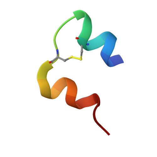
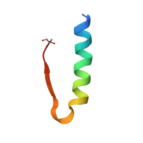
5HPU

Insulin with proline analog HyP at position B28 in the R6 state


Experimental Data Snapshot

  • Method: X-RAY DIFFRACTION
  • Resolution: 2.20 Å
  • R-Value Free: 0.218 
  • R-Value Work: 0.158 
  • R-Value Observed: 0.161 

Starting Model: experimental
View more details

wwPDB Validation   3D Report Full Report


This is version 1.4 of the entry. See complete history


Literature

4S-Hydroxylation of Insulin at ProB28 Accelerates Hexamer Dissociation and Delays Fibrillation.

Lieblich, S.A.Fang, K.Y.Cahn, J.K.B.Rawson, J.LeBon, J.Ku, H.T.Tirrell, D.A.

(2017) J Am Chem Soc 139: 8384-8387

  • DOI: https://doi.org/10.1021/jacs.7b00794
  • Primary Citation of Related Structures:  
    5HPR, 5HPU, 5HQI, 5HRQ

  • PubMed Abstract: 

    Daily injections of insulin provide lifesaving benefits to millions of diabetics. But currently available prandial insulins are suboptimal: The onset of action is delayed by slow dissociation of the insulin hexamer in the subcutaneous space, and insulin forms amyloid fibrils upon storage in solution. Here we show, through the use of noncanonical amino acid mutagenesis, that replacement of the proline residue at position 28 of the insulin B-chain (ProB28) by (4S)-hydroxyproline (Hzp) yields an active form of insulin that dissociates more rapidly, and fibrillates more slowly, than the wild-type protein. Crystal structures of dimeric and hexameric insulin preparations suggest that a hydrogen bond between the hydroxyl group of Hzp and a backbone amide carbonyl positioned across the dimer interface may be responsible for the altered behavior. The effects of hydroxylation are stereospecific; replacement of ProB28 by (4R)-hydroxyproline (Hyp) causes little change in the rates of fibrillation and hexamer disassociation. These results demonstrate a new approach that fuses the concepts of medicinal chemistry and protein design, and paves the way to further engineering of insulin and other therapeutic proteins.


  • Organizational Affiliation

    Division of Chemistry and Chemical Engineering, California Institute of Technology , Pasadena, California 91125, United States.


Macromolecules

Find similar proteins by:  Sequence   |   3D Structure  

Entity ID: 1
MoleculeChains Sequence LengthOrganismDetailsImage
Insulin, chain A
A, C
21Homo sapiensMutation(s): 0 
Gene Names: INS
UniProt & NIH Common Fund Data Resources
Find proteins for P01308 (Homo sapiens)
Explore P01308 
Go to UniProtKB:  P01308
PHAROS:  P01308
GTEx:  ENSG00000254647 
Entity Groups  
Sequence Clusters30% Identity50% Identity70% Identity90% Identity95% Identity100% Identity
UniProt GroupP01308
Sequence Annotations
Expand
  • Reference Sequence
Find similar proteins by:  (by identity cutoff)  |  3D Structure
Entity ID: 2
MoleculeChains Sequence LengthOrganismDetailsImage
Insulin, chain B
B, D
30Homo sapiensMutation(s): 0 
Gene Names: INS
UniProt & NIH Common Fund Data Resources
Find proteins for P01308 (Homo sapiens)
Explore P01308 
Go to UniProtKB:  P01308
PHAROS:  P01308
GTEx:  ENSG00000254647 
Entity Groups  
Sequence Clusters30% Identity50% Identity70% Identity90% Identity95% Identity100% Identity
UniProt GroupP01308
Sequence Annotations
Expand
  • Reference Sequence
Small Molecules
Modified Residues  1 Unique
IDChains TypeFormula2D DiagramParent
HYP
Query on HYP
B, D
L-PEPTIDE LINKINGC5 H9 N O3PRO
Experimental Data & Validation

Experimental Data

  • Method: X-RAY DIFFRACTION
  • Resolution: 2.20 Å
  • R-Value Free: 0.218 
  • R-Value Work: 0.158 
  • R-Value Observed: 0.161 
  • Space Group: H 3
Unit Cell:
Length ( Å )Angle ( ˚ )
a = 77.757α = 90
b = 77.757β = 90
c = 40.397γ = 120
Software Package:
Software NamePurpose
REFMACrefinement
SCALAdata scaling
PHASERphasing
PDB_EXTRACTdata extraction

Structure Validation

View Full Validation Report


Entry History & Funding Information

Deposition Data


Funding OrganizationLocationGrant Number
Novo Nordisk FoundationDenmark--

Revision History  (Full details and data files)

  • Version 1.0: 2017-01-25
    Type: Initial release
  • Version 1.1: 2017-07-12
    Changes: Database references
  • Version 1.2: 2017-11-22
    Changes: Structure summary
  • Version 1.3: 2020-01-15
    Changes: Author supporting evidence
  • Version 1.4: 2023-09-27
    Changes: Data collection, Database references, Derived calculations, Refinement description