5HOY | pdb_00005hoy

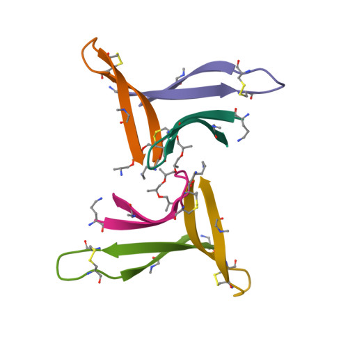
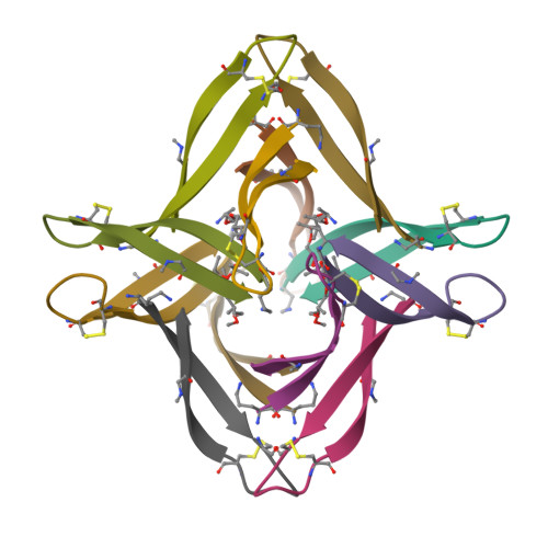
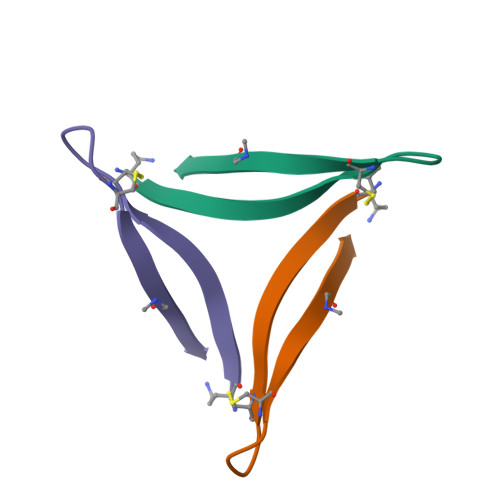
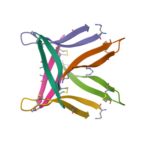
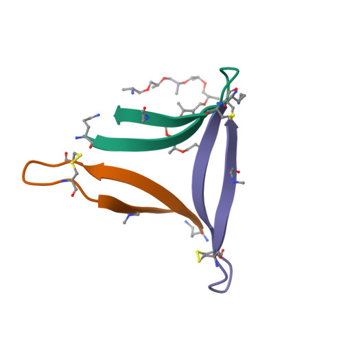
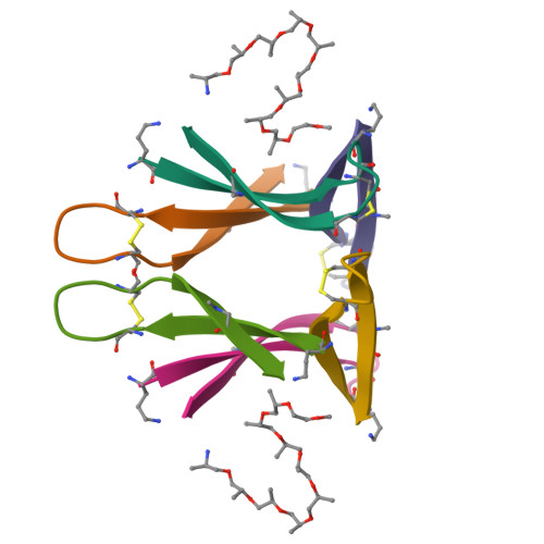
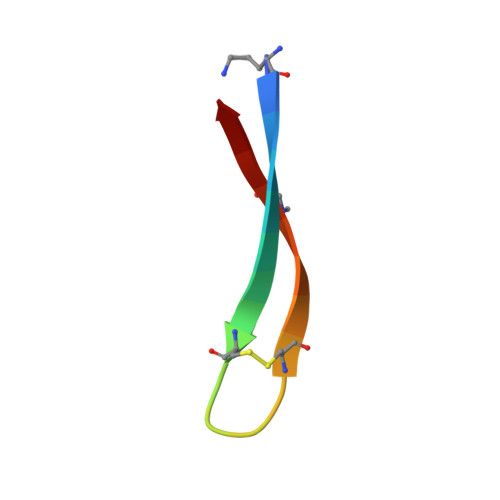
X-ray crystallographic structure of an A-beta 17_36 beta-hairpin. X-ray diffractometer data set. (LVFFAEDCGSNKCAII(SAR)LMV).


Experimental Data Snapshot

  • Method: X-RAY DIFFRACTION
  • Resolution: 2.29 Å
  • R-Value Free: 
    0.276 (Depositor), 0.280 (DCC) 
  • R-Value Work: 
    0.245 (Depositor), 0.250 (DCC) 
  • R-Value Observed: 
    0.248 (Depositor) 

Starting Model: experimental
View more details

wwPDB Validation   3D Report Full Report


Ligand Structure Quality Assessment 

Created with Raphaël 2.3.0Worse 01 BetterLigand structure goodness of fit to experimental dataBest fitted JEFClick on this verticalbar to view details

This is version 1.4 of the entry. See complete history


Literature

X-ray Crystallographic Structures of a Trimer, Dodecamer, and Annular Pore Formed by an A beta 17-36 beta-Hairpin.

Kreutzer, A.G.Hamza, I.L.Spencer, R.K.Nowick, J.S.

(2016) J Am Chem Soc 138: 4634-4642

  • DOI: https://doi.org/10.1021/jacs.6b01332
  • Primary Citation of Related Structures:  
    5HOW, 5HOX, 5HOY

  • PubMed Abstract: 

    High-resolution structures of oligomers formed by the β-amyloid peptide Aβ are needed to understand the molecular basis of Alzheimer's disease and develop therapies. This paper presents the X-ray crystallographic structures of oligomers formed by a 20-residue peptide segment derived from Aβ. The development of a peptide in which Aβ17-36 is stabilized as a β-hairpin is described, and the X-ray crystallographic structures of oligomers it forms are reported. Two covalent constraints act in tandem to stabilize the Aβ17-36 peptide in a hairpin conformation: a δ-linked ornithine turn connecting positions 17 and 36 to create a macrocycle and an intramolecular disulfide linkage between positions 24 and 29. An N-methyl group at position 33 blocks uncontrolled aggregation. The peptide readily crystallizes as a folded β-hairpin, which assembles hierarchically in the crystal lattice. Three β-hairpin monomers assemble to form a triangular trimer, four trimers assemble in a tetrahedral arrangement to form a dodecamer, and five dodecamers pack together to form an annular pore. This hierarchical assembly provides a model, in which full-length Aβ transitions from an unfolded monomer to a folded β-hairpin, which assembles to form oligomers that further pack to form an annular pore. This model may provide a better understanding of the molecular basis of Alzheimer's disease at atomic resolution.


  • Organizational Affiliation

    Department of Chemistry, University of California, Irvine , Irvine, California 92697-2025, United States.


Macromolecules

Find similar proteins by:  Sequence   |   3D Structure  

Entity ID: 1
MoleculeChains Sequence LengthOrganismDetailsImage
Amyloid beta A4 protein
A, B, C, D, E
21Homo sapiensMutation(s): 2 
Membrane Entity: Yes 
UniProt & NIH Common Fund Data Resources
Find proteins for P05067 (Homo sapiens)
Explore P05067 
Go to UniProtKB:  P05067
PHAROS:  P05067
GTEx:  ENSG00000142192 
Entity Groups  
Sequence Clusters30% Identity50% Identity70% Identity90% Identity95% Identity100% Identity
UniProt GroupP05067
Sequence Annotations
Expand
  • Reference Sequence
Small Molecules
Ligands 1 Unique
IDChains Name / Formula / InChI Key2D Diagram3D Interactions
JEF
Query on JEF

Download Ideal Coordinates CCD File 
G [auth D]O-(O-(2-AMINOPROPYL)-O'-(2-METHOXYETHYL)POLYPROPYLENE GLYCOL 500)
C30 H63 N O10
ICCXIDTYQFYPNV-RUMGZKRTSA-N
Modified Residues  2 Unique
IDChains TypeFormula2D DiagramParent
ORN
Query on ORN
A, B, C, D, E
L-PEPTIDE LINKINGC5 H12 N2 O2ALA
SAR
Query on SAR
A, B, C, D, E
PEPTIDE LINKINGC3 H7 N O2GLY
Experimental Data & Validation

Experimental Data

  • Method: X-RAY DIFFRACTION
  • Resolution: 2.29 Å
  • R-Value Free:  0.276 (Depositor), 0.280 (DCC) 
  • R-Value Work:  0.245 (Depositor), 0.250 (DCC) 
  • R-Value Observed: 0.248 (Depositor) 
Space Group: P 61 2 2
Unit Cell:
Length ( Å )Angle ( ˚ )
a = 97.649α = 90
b = 97.649β = 90
c = 97.781γ = 120
Software Package:
Software NamePurpose
PHENIXrefinement
MOSFLMdata reduction
Aimlessdata scaling
PHASERphasing

Structure Validation

View Full Validation Report


Ligand Structure Quality Assessment 

Created with Raphaël 2.3.0Worse 01 BetterLigand structure goodness of fit to experimental dataBest fitted JEFClick on this verticalbar to view details

Entry History & Funding Information

Deposition Data


Funding OrganizationLocationGrant Number
National Institutes of Health/National Institute of General Medical Sciences (NIH/NIGMS)United StatesGM097562

Revision History  (Full details and data files)

  • Version 1.0: 2016-03-23
    Type: Initial release
  • Version 1.1: 2016-04-20
    Changes: Database references
  • Version 1.2: 2017-09-13
    Changes: Author supporting evidence, Derived calculations
  • Version 1.3: 2019-12-25
    Changes: Author supporting evidence
  • Version 1.4: 2023-09-27
    Changes: Data collection, Database references, Derived calculations, Refinement description