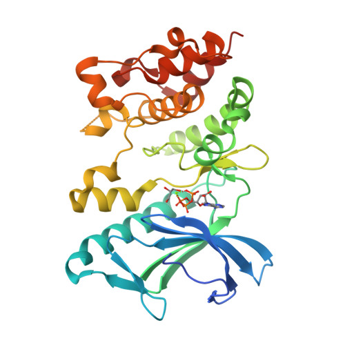
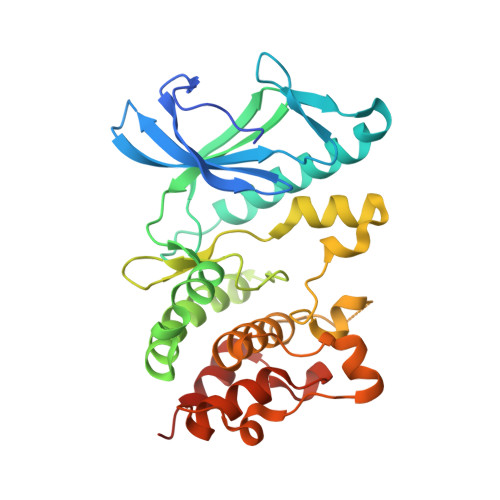
5HNV

Crystal structure of PpkA


Experimental Data Snapshot

  • Method: X-RAY DIFFRACTION
  • Resolution: 1.41 Å
  • R-Value Free: 0.210 
  • R-Value Work: 0.183 
  • R-Value Observed: 0.184 

wwPDB Validation   3D Report Full Report


Ligand Structure Quality Assessment 


This is version 1.4 of the entry. See complete history


Literature

Crystal structures of the kinase domain of PpkA, a key regulatory component of T6SS, reveal a general inhibitory mechanism.

Li, P.P.Xu, D.Q.Ma, T.Q.Wang, D.Y.Li, W.D.He, J.H.Ran, T.T.Wang, W.W.

(2018) Biochem J 475: 2209-2224

  • DOI: https://doi.org/10.1042/BCJ20180077
  • Primary Citation of Related Structures:  
    5HNV, 5X1Q, 5X1R, 5X1S, 5X1T

  • PubMed Abstract: 

    The type VI secretion system (T6SS) is a versatile and widespread export system found in many Gram-negative bacteria that delivers effector proteins into target cells. The functions of T6SSs are tightly regulated by diverse mechanisms at multiple levels, including post-translational modification through threonine phosphorylation via the Ser/Thr protein kinase (STPK) PpkA. Here, we identified that PpkA is essential for T6SS secretion in Serratia marcescens since its deletion eliminated the secretion of haemolysin co-regulated protein, while the periplasmic and transmembrane portion of PpkA was found to be disposable for T6SS secretion. We further determined the crystal structure of the kinase domain of PpkA (PpkA-294). The structure of PpkA-294 was determined in its apo form to a 1.6 Å resolution as well as in complex with ATP to a 1.41 Å resolution and with an ATP analogue AMP-PCP to a 1.45 Å resolution. The residues in the activation loop of PpkA-294 were fully determined, and the N-terminus of the loop was folded into an unprecedented inhibitory helix, revealing that the PpkA kinase domain was in an auto-inhibitory state. The ternary MgATP-PpkA-294 complex was also inactive with nucleotide ribose and phosphates in unexpected and unproductive conformations. The αC-helix in the inactive PpkA-294 adopted a conformation towards the active site but with the conserved glutamate in the helix rotated away, which we suggest to be a general conformation for all STPK kinases in the inactive form. Structural comparison of PpkA with its eukaryotic homologues reinforced the universal regulation mechanism of protein kinases.


  • Organizational Affiliation

    Department of Microbiology, College of Life Sciences, Nanjing Agricultural University, 210095 Nanjing, PR China.


Macromolecules
Find similar proteins by:  (by identity cutoff)  |  3D Structure
Entity ID: 1
MoleculeChains Sequence LengthOrganismDetailsImage
PpkA N terminal302Serratia sp. FS14Mutation(s): 0 
UniProt
Find proteins for A0A1S4NYE5 (Serratia sp. (strain FS14))
Explore A0A1S4NYE5 
Go to UniProtKB:  A0A1S4NYE5
Entity Groups  
Sequence Clusters30% Identity50% Identity70% Identity90% Identity95% Identity100% Identity
UniProt GroupA0A1S4NYE5
Sequence Annotations
Expand
  • Reference Sequence
Small Molecules
Ligands 3 Unique
IDChains Name / Formula / InChI Key2D Diagram3D Interactions
ATP
Query on ATP

Download Ideal Coordinates CCD File 
B [auth A]ADENOSINE-5'-TRIPHOSPHATE
C10 H16 N5 O13 P3
ZKHQWZAMYRWXGA-KQYNXXCUSA-N
GOL
Query on GOL

Download Ideal Coordinates CCD File 
D [auth A]GLYCEROL
C3 H8 O3
PEDCQBHIVMGVHV-UHFFFAOYSA-N
MG
Query on MG

Download Ideal Coordinates CCD File 
C [auth A]MAGNESIUM ION
Mg
JLVVSXFLKOJNIY-UHFFFAOYSA-N
Experimental Data & Validation

Experimental Data

  • Method: X-RAY DIFFRACTION
  • Resolution: 1.41 Å
  • R-Value Free: 0.210 
  • R-Value Work: 0.183 
  • R-Value Observed: 0.184 
  • Space Group: P 21 21 2
Unit Cell:
Length ( Å )Angle ( ˚ )
a = 65.753α = 90
b = 78.282β = 90
c = 59.162γ = 90
Software Package:
Software NamePurpose
PHENIXrefinement
XDSdata reduction
SCALAdata scaling
PHASERphasing

Structure Validation

View Full Validation Report


Ligand Structure Quality Assessment 


Entry History & Funding Information

Deposition Data


Funding OrganizationLocationGrant Number
National Natural Science Foundation of ChinaChina31170686
National Natural Science Foundation of ChinaChina31400055
National Natural Science Foundation of ChinaChina31100028
the Natural Science Foundation of Jiangsu ProvinceChinaBK20140690

Revision History  (Full details and data files)

  • Version 1.0: 2017-01-25
    Type: Initial release
  • Version 1.1: 2017-09-27
    Changes: Data collection
  • Version 1.2: 2017-10-18
    Changes: Author supporting evidence
  • Version 1.3: 2019-08-21
    Changes: Data collection, Database references
  • Version 1.4: 2024-03-20
    Changes: Data collection, Database references