Discovery of Novel 3,3-Disubstituted Piperidines as Orally Bioavailable, Potent, and Efficacious HDM2-p53 Inhibitors.
Bogen, S.L., Pan, W., Gibeau, C.R., Lahue, B.R., Ma, Y., Nair, L.G., Seigel, E., Shipps, G.W., Tian, Y., Wang, Y., Lin, Y., Liu, M., Liu, S., Mirza, A., Wang, X., Lipari, P., Seidel-Dugan, C., Hicklin, D.J., Bishop, W.R., Rindgen, D., Nomeir, A., Prosise, W., Reichert, P., Scapin, G., Strickland, C., Doll, R.J.(2016) ACS Med Chem Lett 7: 324-329
- PubMed: 26985323 Search on PubMedSearch on PubMed Central
- DOI: https://doi.org/10.1021/acsmedchemlett.5b00472
- Primary Citation Related Structures:
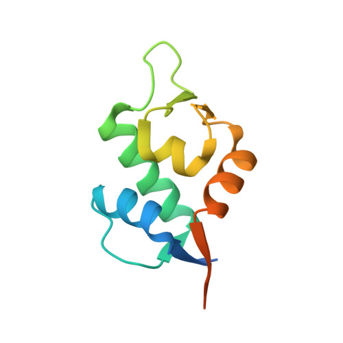
5HMH, 5HMI, 5HMK - PubMed Abstract:
A new subseries of substituted piperidines as p53-HDM2 inhibitors exemplified by 21 has been developed from the initial lead 1. Research focused on optimization of a crucial HDM2 Trp23-ligand interaction led to the identification of 2-(trifluoromethyl)thiophene as the preferred moiety. Further investigation of the Leu26 pocket resulted in potent, novel substituted piperidine inhibitors of the HDM2-p53 interaction that demonstrated tumor regression in several human cancer xenograft models in mice. The structure of HDM2 in complex with inhibitors 3, 10, and 21 is described.
- Discovery Chemistry, Merck Research Laboratories , Kenilworth, New Jersey 07033, United States.
Organizational Affiliation: