5HLI | pdb_00005hli

Structure of Disulfide formed AbfR


Experimental Data Snapshot

  • Method: X-RAY DIFFRACTION
  • Resolution: 2.05 Å
  • R-Value Free: 
    0.245 (Depositor), 0.246 (DCC) 
  • R-Value Work: 
    0.212 (Depositor), 0.213 (DCC) 
  • R-Value Observed: 
    0.214 (Depositor) 

Starting Model: experimental
View more details

wwPDB Validation 3D Report Full Report

Validation slider image for 5HLI

This is version 1.3 of the entry. See complete history

Literature

Structural Insights into the Redox-Sensing Mechanism of MarR-Type Regulator AbfR.

Liu, G.Liu, X.Xu, H.Liu, X.Zhou, H.Huang, Z.Gan, J.Chen, H.Lan, L.Yang, C.G.

(2017) J Am Chem Soc 139: 1598-1608

  • DOI: https://doi.org/10.1021/jacs.6b11438
  • Primary Citation Related Structures: 
    5HLG, 5HLH, 5HLI

  • PubMed Abstract: 

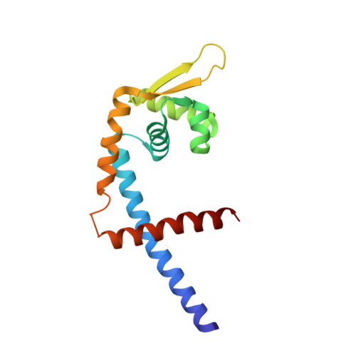
    As a master redox-sensing MarR-family transcriptional regulator, AbfR participates in oxidative stress responses and virulence regulations in Staphylococcus epidermidis. Here, we present structural insights into the DNA-binding mechanism of AbfR in different oxidation states by determining the X-ray crystal structures of a reduced-AbfR/DNA complex, an overoxidized (Cys13-SO 2 H and Cys13-SO 3 H) AbfR/DNA, and 2-disulfide cross-linked AbfR dimer. Together with biochemical analyses, our results suggest that the redox regulation of AbfR-sensing displays two novel features: (i) the reversible disulfide modification, but not the irreversible overoxidation, significantly abolishes the DNA-binding ability of the AbfR repressor; (ii) either 1-disulfide cross-linked or 2-disulfide cross-linked AbfR dimer is biologically significant. The overoxidized species of AbfR, resembling the reduced AbfR in conformation and retaining the DNA-binding ability, does not exist in biologically significant concentrations, however. The 1-disulfide cross-linked modification endows AbfR with significantly weakened capability for DNA-binding. The 2-disulfide cross-linked AbfR adopts a very "open" conformation that is incompatible with DNA-binding. Overall, the concise oxidation chemistry of the redox-active cysteine allows AbfR to sense and respond to oxidative stress correctly and efficiently.


  • Organizational Affiliation
    • Laboratory of Chemical Biology, State Key Laboratory of Drug Research, Shanghai Institute of Materia Medica, Chinese Academy of Sciences , Shanghai 201203, China.

Macromolecule Content 

  • Total Structure Weight: 35.5 kDa 
  • Atom Count: 2,425 
  • Modeled Residue Count: 282 
  • Deposited Residue Count: 298 
  • Unique protein chains: 1

Macromolecules

Find similar proteins by:|  3D Structure
Entity ID: 1
MoleculeChains  Sequence LengthOrganismDetailsImage
MarR family transcriptional regulator
A, B
149Staphylococcus epidermidisMutation(s): 1 
Gene Names: ADT70_02765
UniProt
Find proteins for Q5HKZ1 (Staphylococcus epidermidis (strain ATCC 35984 / DSM 28319 / BCRC 17069 / CCUG 31568 / BM 3577 / RP62A))
Explore Q5HKZ1 
Go to UniProtKB:  Q5HKZ1
Entity Groups
Sequence Clusters30% Identity50% Identity70% Identity90% Identity95% Identity100% Identity
UniProt GroupQ5HKZ1
Sequence Annotations
Expand
Reference Sequence

Experimental Data & Validation

Experimental Data

  • Method: X-RAY DIFFRACTION
  • Resolution: 2.05 Å
  • R-Value Free:  0.245 (Depositor), 0.246 (DCC) 
  • R-Value Work:  0.212 (Depositor), 0.213 (DCC) 
  • R-Value Observed: 0.214 (Depositor) 
Space Group: P 1 21 1
Unit Cell:
Length ( Å )Angle ( ˚ )
a = 46.534α = 90
b = 51.686β = 103.73
c = 85.935γ = 90
Software Package:
Software NamePurpose
PHENIXrefinement
HKL-2000data reduction
HKL-2000data scaling
PHASERphasing

Structure Validation

View Full Validation Report


Entry History 

Deposition Data

Revision History  (Full details and data files)

  • Version 1.0: 2017-01-25
    Type: Initial release
  • Version 1.1: 2017-04-05
    Changes: Database references
  • Version 1.2: 2023-11-08
    Changes: Data collection, Database references, Derived calculations, Refinement description
  • Version 1.3: 2024-10-23
    Changes: Structure summary