Crystal structure and interaction studies of the human FBxo3 ApaG domain.
Krzysiak, T.C., Chen, B.B., Lear, T., Mallampalli, R.K., Gronenborn, A.M.(2016) FEBS J 283: 2091-2101
- PubMed: 27010866
- DOI: https://doi.org/10.1111/febs.13721
- Primary Citation of Related Structures:
5HDW - PubMed Abstract:
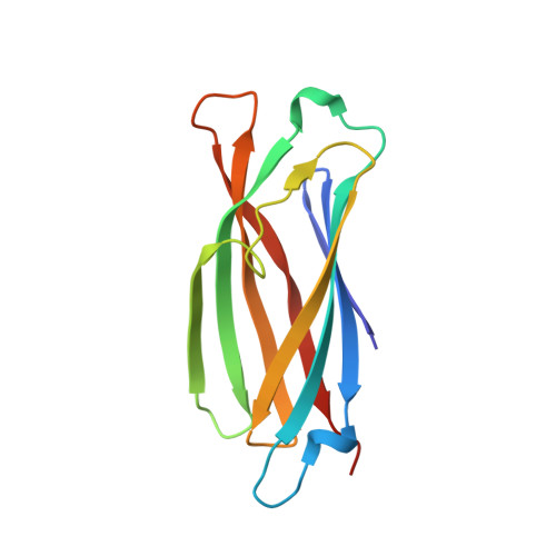
Transcriptional activation of proinflammatory cytokines, mediated by tumor necrosis factor receptor-associated factors (TRAFs), is in part triggered by the degradation of the F-box protein, FBxl2, via an E3 ligase that contains another F-box protein, FBxo3. The ApaG domain of FBxo3 is required for the interaction with and degradation of FBxl2 [Mallampalli RK et al., (2013) J Immunol 191, 5247-5255]. Here, we report the X-ray structure of the human FBxo3 ApaG domain, residues 278-407, at 2.0 Å resolution. Like bacterial ApaG proteins, this domain is characterized by a classic Immunoglobin/Fibronectin III-type fold, comprising a seven-stranded β-sheet core, surrounded by four extended loops. Although cation binding had been proposed for bacterial ApaG proteins, no interactions with Mg(2+) or Co(2+) were detected for the human ApaG domain. In addition, dinucleotide polyphosphates, which have been reported to be second messengers in the inflammation response and targets of the bacterial apaG-containing operon, are not bound by the human ApaG domain. In the context of the full-length protein, loop 1, comprising residues 294-303, is critical for the interaction with FBxl2. However, titration of the individual ApaG domain with a 15-mer FBxl2 peptide that was phosphorylated on the crucial T404, as well as the inability of the ApaG domain to interact with full-length FBxl2, assessed by coimmunoprecipitation, indicate that the ApaG domain alone is necessary, but not sufficient for binding and degradation of FBxl2. PDB ID (5HDW).
Organizational Affiliation:
Department of Structural Biology, Pittsburgh Center for HIV Protein Interactions, University of Pittsburgh School of Medicine, PA, USA.


