5HBN | pdb_00005hbn

ClpC N-terminal domain with bound phospho-arginine


Experimental Data Snapshot

  • Method: X-RAY DIFFRACTION
  • Resolution: 1.60 Å
  • R-Value Free: 
    0.166 (Depositor), 0.167 (DCC) 
  • R-Value Work: 
    0.145 (Depositor), 0.147 (DCC) 
  • R-Value Observed: 
    0.147 (Depositor) 

Starting Model: experimental
View more details

wwPDB Validation 3D Report Full Report

Validation slider image for 5HBN

Ligand Structure Quality Assessment 


This is version 2.1 of the entry. See complete history

Literature

Arginine phosphorylation marks proteins for degradation by a Clp protease.

Trentini, D.B.Suskiewicz, M.J.Heuck, A.Kurzbauer, R.Deszcz, L.Mechtler, K.Clausen, T.

(2016) Nature 539: 48-53

  • DOI: https://doi.org/10.1038/nature20122
  • Primary Citation Related Structures: 
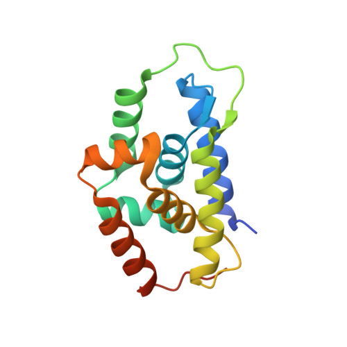
    5HBN

  • PubMed Abstract: 

    Protein turnover is a tightly controlled process that is crucial for the removal of aberrant polypeptides and for cellular signalling. Whereas ubiquitin marks eukaryotic proteins for proteasomal degradation, a general tagging system for the equivalent bacterial Clp proteases is not known. Here we describe the targeting mechanism of the ClpC-ClpP proteolytic complex from Bacillus subtilis. Quantitative affinity proteomics using a ClpP-trapping mutant show that proteins phosphorylated on arginine residues are selectively targeted to ClpC-ClpP. In vitro reconstitution experiments demonstrate that arginine phosphorylation by the McsB kinase is required and sufficient for the degradation of substrate proteins. The docking site for phosphoarginine is located in the amino-terminal domain of the ClpC ATPase, as resolved at high resolution in a co-crystal structure. Together, our data demonstrate that phosphoarginine functions as a bona fide degradation tag for the ClpC-ClpP protease. This system, which is widely distributed across Gram-positive bacteria, is functionally analogous to the eukaryotic ubiquitin-proteasome system.


  • Organizational Affiliation
    • Research Institute of Molecular Pathology (IMP), Dr-Bohr-Gasse 7, 1030 Vienna, Austria.

Macromolecule Content 

  • Total Structure Weight: 18.19 kDa 
  • Atom Count: 1,264 
  • Modeled Residue Count: 145 
  • Deposited Residue Count: 156 
  • Unique protein chains: 1

Macromolecules

Find similar proteins by:|  3D Structure
Entity ID: 1
MoleculeChains  Sequence LengthOrganismDetailsImage
Negative regulator of genetic competence ClpC/MecB156Bacillus subtilis subsp. subtilis str. 168Mutation(s): 0 
Gene Names: clpCmecBBSU00860
UniProt
Find proteins for P37571 (Bacillus subtilis (strain 168))
Explore P37571 
Go to UniProtKB:  P37571
Entity Groups
Sequence Clusters30% Identity50% Identity70% Identity90% Identity95% Identity100% Identity
UniProt GroupP37571
Sequence Annotations
Expand
Reference Sequence

Small Molecules

Ligands 3 Unique
IDChains Name / Formula / InChI Key2D Diagram3D Interactions
RPI

Query on RPI


Download:Ideal Coordinates CCD File
G [auth A],
H [auth A]
phospho-arginine
C6 H15 N4 O5 P
CCTIOCVIZPCTGO-BYPYZUCNSA-N
SO4

Query on SO4


Download:Ideal Coordinates CCD File
B [auth A],
C [auth A],
D [auth A],
E [auth A],
F [auth A]
SULFATE ION
O4 S
QAOWNCQODCNURD-UHFFFAOYSA-L
ACT

Query on ACT


Download:Ideal Coordinates CCD File
I [auth A]ACETATE ION
C2 H3 O2
QTBSBXVTEAMEQO-UHFFFAOYSA-M

Experimental Data & Validation

Experimental Data

  • Method: X-RAY DIFFRACTION
  • Resolution: 1.60 Å
  • R-Value Free:  0.166 (Depositor), 0.167 (DCC) 
  • R-Value Work:  0.145 (Depositor), 0.147 (DCC) 
  • R-Value Observed: 0.147 (Depositor) 
Space Group: P 65
Unit Cell:
Length ( Å )Angle ( ˚ )
a = 84.6α = 90
b = 84.6β = 90
c = 29.85γ = 120
Software Package:
Software NamePurpose
PHENIXrefinement
XDSdata reduction
XSCALEdata scaling
PHASERphasing

Structure Validation

View Full Validation Report


Ligand Structure Quality Assessment 


Entry History 

Deposition Data

Revision History  (Full details and data files)

  • Version 1.0: 2016-10-12
    Type: Initial release
  • Version 1.1: 2016-10-26
    Changes: Database references
  • Version 1.2: 2016-11-09
    Changes: Database references
  • Version 1.3: 2019-07-17
    Changes: Data collection
  • Version 1.4: 2019-10-16
    Changes: Data collection
  • Version 2.0: 2023-11-15
    Changes: Advisory, Atomic model, Data collection, Database references
  • Version 2.1: 2024-01-10
    Changes: Refinement description