5H9Y

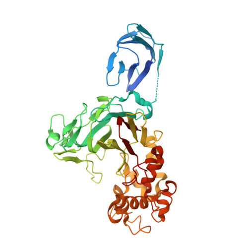
Crystal structure of GH family 64 laminaripentaose-producing beta-1,3-glucanase from Paenibacillus barengoltzii complexed with laminarihexaose.


Experimental Data Snapshot

  • Method: X-RAY DIFFRACTION
  • Resolution: 1.97 Å
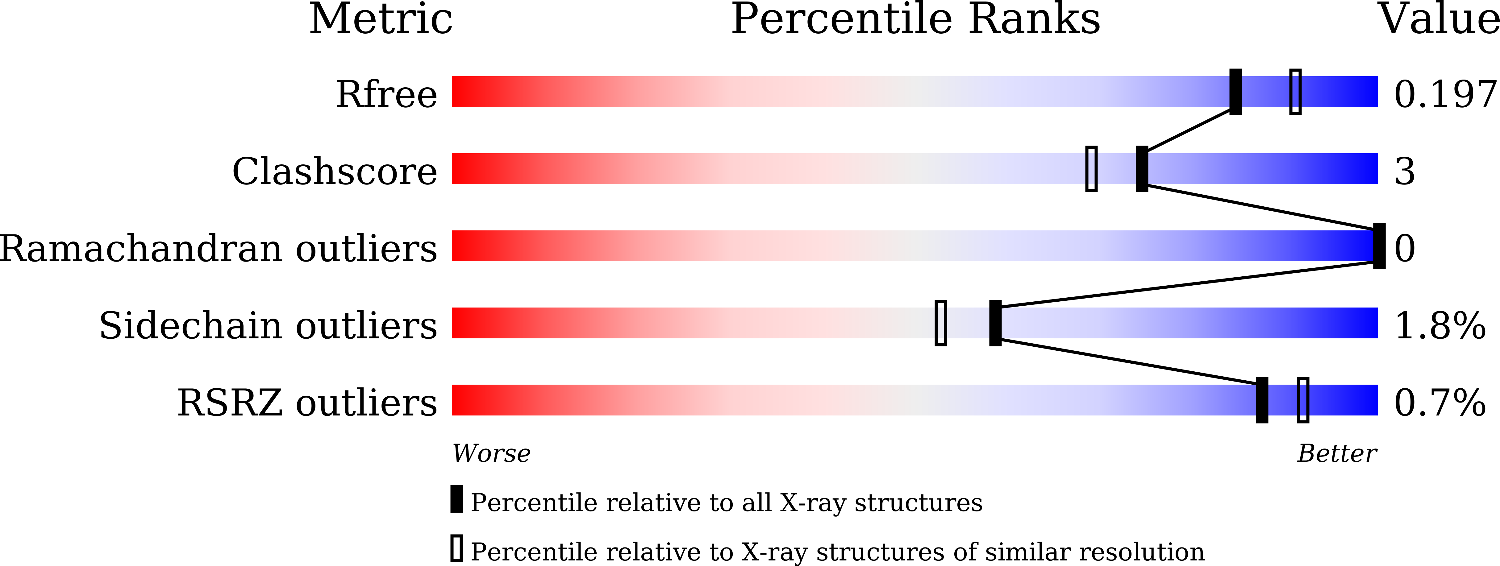
  • R-Value Free: 0.197 
  • R-Value Work: 0.167 
  • R-Value Observed: 0.169 

Starting Model: experimental
View more details

wwPDB Validation   3D Report Full Report


This is version 2.1 of the entry. See complete history


Literature

The recognition mechanism of triple-helical beta-1,3-glucan by a beta-1,3-glucanase

Qin, Z.Yang, D.You, X.Liu, Y.Hu, S.Yan, Q.Yang, S.Jiang, Z.

(2017) Chem Commun (Camb) 53: 9368-9371

  • DOI: https://doi.org/10.1039/c7cc03330c
  • Primary Citation of Related Structures:  
    5H9X, 5H9Y

  • PubMed Abstract: 

    β-1,3-Glucan is one of the most abundant polysaccharides in fungi. Recognition of β-1,3-glucan occurs in both hydrolysis by glycoside hydrolases and immunological recognition. Our study provides a novel structural account of how glycoside hydrolase recognizes and hydrolyzes substrates in a triple-helical form and presents a general structural basis of β-1,3-glucan recognition.


  • Organizational Affiliation

    Beijing Advanced Innovation Center for Food Nutrition and Human Health, College of Food Science and Nutritional Engineering, China Agricultural University, Beijing 100083, China. zhqjiang@cau.edu.cn ysq@cau.edu.cn.


Macromolecules
Find similar proteins by:  (by identity cutoff)  |  3D Structure
Entity ID: 1
MoleculeChains Sequence LengthOrganismDetailsImage
beta-1,3-glucanase444Paenibacillus barengoltziiMutation(s): 0 
EC: 3.2.1.39
UniProt
Find proteins for A0A1S4NYE2 (Paenibacillus barengoltzii)
Explore A0A1S4NYE2 
Go to UniProtKB:  A0A1S4NYE2
Entity Groups  
Sequence Clusters30% Identity50% Identity70% Identity90% Identity95% Identity100% Identity
UniProt GroupA0A1S4NYE2
Sequence Annotations
Expand
  • Reference Sequence
Oligosaccharides

Help

Entity ID: 2
MoleculeChains Length2D Diagram Glycosylation3D Interactions
beta-D-glucopyranose-(1-3)-beta-D-glucopyranose-(1-3)-beta-D-glucopyranose-(1-3)-beta-D-glucopyranose-(1-3)-beta-D-glucopyranose
B
5N/A
Glycosylation Resources
GlyTouCan:  G03330ED
GlyCosmos:  G03330ED
Entity ID: 3
MoleculeChains Length2D Diagram Glycosylation3D Interactions
beta-D-glucopyranose-(1-3)-beta-D-glucopyranose-(1-3)-beta-D-glucopyranose-(1-3)-beta-D-glucopyranose
C
4N/A
Glycosylation Resources
GlyTouCan:  G87841AX
GlyCosmos:  G87841AX
GlyGen:  G87841AX
Small Molecules
Ligands 1 Unique
IDChains Name / Formula / InChI Key2D Diagram3D Interactions
TLA
Query on TLA

Download Ideal Coordinates CCD File 
D [auth A]L(+)-TARTARIC ACID
C4 H6 O6
FEWJPZIEWOKRBE-JCYAYHJZSA-N
Experimental Data & Validation

Experimental Data

  • Method: X-RAY DIFFRACTION
  • Resolution: 1.97 Å
  • R-Value Free: 0.197 
  • R-Value Work: 0.167 
  • R-Value Observed: 0.169 
  • Space Group: C 1 2 1
Unit Cell:
Length ( Å )Angle ( ˚ )
a = 132.561α = 90
b = 65.191β = 104.04
c = 61.426γ = 90
Software Package:
Software NamePurpose
PHENIXrefinement
HKL-2000data reduction
HKL-2000data scaling
PHENIXphasing
PHENIXmodel building

Structure Validation

View Full Validation Report


Entry History 

Deposition Data

Revision History  (Full details and data files)

  • Version 1.0: 2017-02-15
    Type: Initial release
  • Version 1.1: 2017-09-06
    Changes: Database references
  • Version 1.2: 2017-09-27
    Changes: Data collection
  • Version 2.0: 2020-07-29
    Type: Remediation
    Reason: Carbohydrate remediation
    Changes: Advisory, Atomic model, Data collection, Derived calculations, Refinement description, Structure summary
  • Version 2.1: 2023-11-08
    Changes: Data collection, Database references, Derived calculations, Refinement description, Structure summary