5H8V

Crystal structure of the complex between maize Sulfite Reductase and ferredoxin in the form-1 crystal


Experimental Data Snapshot

  • Method: X-RAY DIFFRACTION
  • Resolution: 2.20 Å
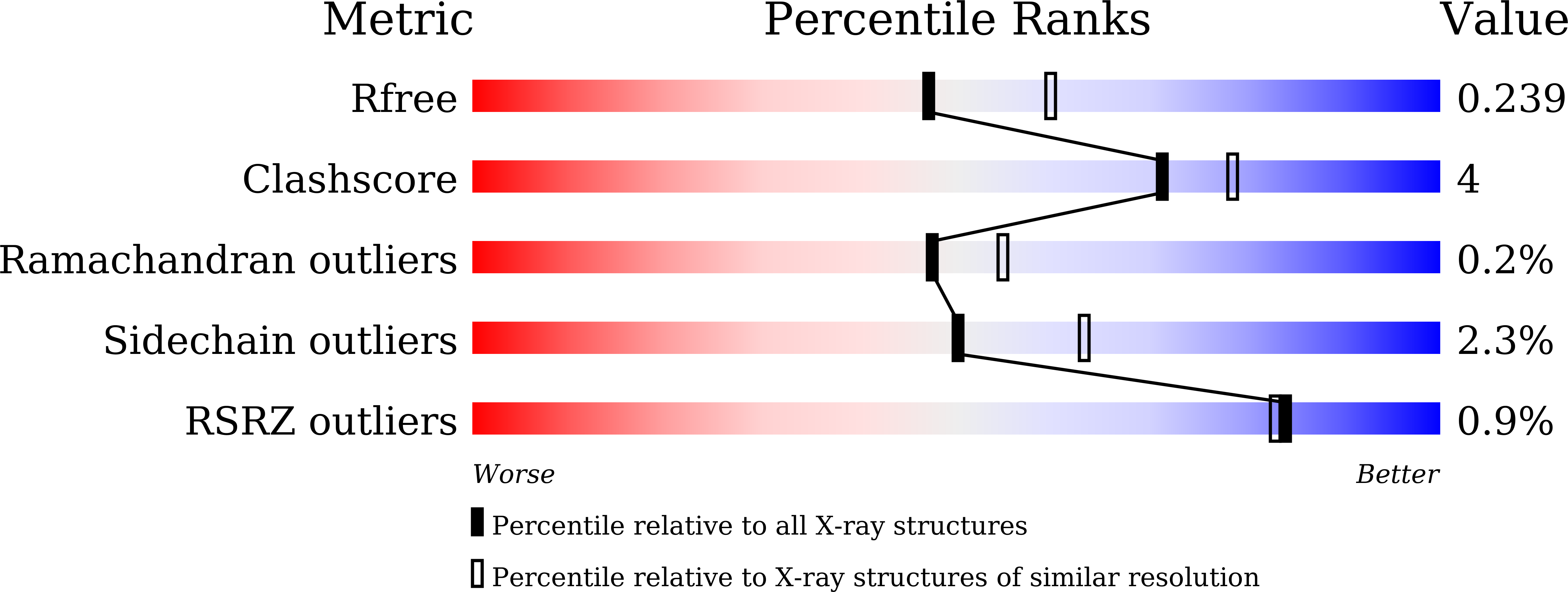
  • R-Value Free: 0.239 
  • R-Value Work: 0.200 
  • R-Value Observed: 0.201 

wwPDB Validation   3D Report Full Report


Ligand Structure Quality Assessment 


This is version 2.1 of the entry. See complete history


Literature

Structural and mutational studies of an electron transfer complex of maize sulfite reductase and ferredoxin.

Kim, J.Y.Nakayama, M.Toyota, H.Kurisu, G.Hase, T.

(2016) J Biochem 160: 101-109

  • DOI: https://doi.org/10.1093/jb/mvw016
  • Primary Citation of Related Structures:  
    5H8V, 5H8Y, 5H92

  • PubMed Abstract: 

    The structure of the complex of maize sulfite reductase (SiR) and ferredoxin (Fd) has been determined by X-ray crystallography. Co-crystals of the two proteins prepared under different conditions were subjected to the diffraction analysis and three possible structures of the complex were solved. Although topological relationship of SiR and Fd varied in each of the structures, two characteristics common to all structures were found in the pattern of protein-protein interactions and positional arrangements of redox centres; (i) a few negative residues of Fd contact with a narrow area of SiR with positive electrostatic surface potential and (ii) [2Fe-2S] cluster of Fd and [4Fe-4S] cluster of SiR are in a close proximity with the shortest distance around 12 Å. Mutational analysis of a total of seven basic residues of SiR distributed widely at the interface of the complex showed their importance for supporting an efficient Fd-dependent activity and a strong physical binding to Fd. These combined results suggest that the productive electron transfer complex of SiR and Fd could be formed through multiple processes of the electrostatic intermolecular interaction and this implication is discussed in terms of the multi-functionality of Fd in various redox metabolisms.


  • Organizational Affiliation

    Division of Protein Chemistry and.


Macromolecules
Find similar proteins by:  (by identity cutoff)  |  3D Structure
Entity ID: 1
MoleculeChains Sequence LengthOrganismDetailsImage
Sulfite reductase [ferredoxin], chloroplastic
A, B
583Zea maysMutation(s): 0 
Gene Names: SIR
EC: 1.8.7.1
UniProt
Find proteins for O23813 (Zea mays)
Explore O23813 
Go to UniProtKB:  O23813
Entity Groups  
Sequence Clusters30% Identity50% Identity70% Identity90% Identity95% Identity100% Identity
UniProt GroupO23813
Sequence Annotations
Expand
  • Reference Sequence
Small Molecules
Ligands 4 Unique
IDChains Name / Formula / InChI Key2D Diagram3D Interactions
SRM
Query on SRM

Download Ideal Coordinates CCD File 
D [auth A],
I [auth B]
SIROHEME
C42 H44 Fe N4 O16
DLKSSIHHLYNIKN-MWBYXLBFSA-L
SF4
Query on SF4

Download Ideal Coordinates CCD File 
C [auth A],
H [auth B]
IRON/SULFUR CLUSTER
Fe4 S4
LJBDFODJNLIPKO-UHFFFAOYSA-N
PO4
Query on PO4

Download Ideal Coordinates CCD File 
E [auth A],
J [auth B]
PHOSPHATE ION
O4 P
NBIIXXVUZAFLBC-UHFFFAOYSA-K
MG
Query on MG

Download Ideal Coordinates CCD File 
F [auth A],
G [auth A],
K [auth B],
L [auth B]
MAGNESIUM ION
Mg
JLVVSXFLKOJNIY-UHFFFAOYSA-N
Experimental Data & Validation

Experimental Data

  • Method: X-RAY DIFFRACTION
  • Resolution: 2.20 Å
  • R-Value Free: 0.239 
  • R-Value Work: 0.200 
  • R-Value Observed: 0.201 
  • Space Group: P 41 21 2
Unit Cell:
Length ( Å )Angle ( ˚ )
a = 103.346α = 90
b = 103.346β = 90
c = 255.048γ = 90
Software Package:
Software NamePurpose
REFMACrefinement
HKL-2000data reduction
HKL-2000data scaling
SHARPphasing

Structure Validation

View Full Validation Report


Ligand Structure Quality Assessment 


Entry History & Funding Information

Deposition Data


Funding OrganizationLocationGrant Number
Japan Science and Technology AgencyJapanCREST
Cabinet office of JapanJapanGS016

Revision History  (Full details and data files)

  • Version 1.0: 2016-04-13
    Type: Initial release
  • Version 1.1: 2016-08-10
    Changes: Database references
  • Version 1.2: 2020-02-19
    Changes: Data collection, Derived calculations
  • Version 2.0: 2023-12-27
    Changes: Data collection, Database references, Derived calculations, Non-polymer description, Refinement description, Structure summary
  • Version 2.1: 2024-10-23
    Changes: Structure summary