5H7E | pdb_00005h7e

Crystal Structure of native drCPDase


Experimental Data Snapshot

  • Method: X-RAY DIFFRACTION
  • Resolution: 1.60 Å
  • R-Value Free: 
    0.196 (Depositor), 0.200 (DCC) 
  • R-Value Work: 
    0.175 (Depositor), 0.176 (DCC) 
  • R-Value Observed: 
    0.176 (Depositor) 

Starting Model: experimental
View more details

wwPDB Validation 3D Report Full Report

Validation slider image for 5H7E

This is version 1.1 of the entry. See complete history

Literature

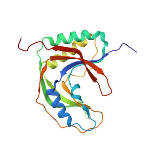
Crystal structure of the RNA 2',3'-cyclic phosphodiesterase from Deinococcus radiodurans

Han, W.Cheng, J.Zhou, C.Hua, Y.Zhao, Y.

(2017) Acta Crystallogr F Struct Biol Commun 73: 276-280

  • DOI: https://doi.org/10.1107/S2053230X17004964
  • Primary Citation Related Structures: 
    5H7E

  • PubMed Abstract: 

    2',3'-Cyclic phosphodiesterase (CPDase) homologues have been found in all domains of life and are involved in diverse RNA and nucleotide metabolisms. The CPDase from Deinococcus radiodurans was crystallized and the crystals diffracted to 1.6 Å resolution, which is the highest resolution currently known for a CPDase structure. Structural comparisons revealed that the enzyme is in an open conformation in the absence of substrate. Nevertheless, the active site is well formed, and the representative motifs interact with sulfate ion, which suggests a conserved catalytic mechanism.


  • Organizational Affiliation
    • Key Laboratory of Chinese Ministry of Agriculture for Nuclear-Agricultural Sciences, Institute of Nuclear-Agricultural Sciences, Zhejiang University, People's Republic of China.

Macromolecule Content 

  • Total Structure Weight: 21.12 kDa 
  • Atom Count: 1,560 
  • Modeled Residue Count: 182 
  • Deposited Residue Count: 182 
  • Unique protein chains: 1

Macromolecules

Find similar proteins by:|  3D Structure
Entity ID: 1
MoleculeChains  Sequence LengthOrganismDetailsImage
RNA 2',3'-cyclic phosphodiesterase182Deinococcus radioduransMutation(s): 0 
Gene Names: DR_2339
EC: 3.1.4 (PDB Primary Data), 3.1.4.58 (UniProt)
UniProt
Find proteins for O32509 (Deinococcus radiodurans)
Explore O32509 
Go to UniProtKB:  O32509
Entity Groups
Sequence Clusters30% Identity50% Identity70% Identity90% Identity95% Identity100% Identity
UniProt GroupO32509
Sequence Annotations
Expand
Reference Sequence

Experimental Data & Validation

Experimental Data

  • Method: X-RAY DIFFRACTION
  • Resolution: 1.60 Å
  • R-Value Free:  0.196 (Depositor), 0.200 (DCC) 
  • R-Value Work:  0.175 (Depositor), 0.176 (DCC) 
  • R-Value Observed: 0.176 (Depositor) 
Space Group: H 3 2
Unit Cell:
Length ( Å )Angle ( ˚ )
a = 91.22α = 90
b = 91.22β = 90
c = 169.83γ = 120
Software Package:
Software NamePurpose
PHENIXrefinement
XSCALEdata scaling
PDB_EXTRACTdata extraction
XDSdata reduction
MOLREPphasing

Structure Validation

View Full Validation Report


Entry History 

Deposition Data

Revision History  (Full details and data files)

  • Version 1.0: 2017-09-27
    Type: Initial release
  • Version 1.1: 2023-11-08
    Changes: Data collection, Database references, Refinement description