5H60 | pdb_00005h60

Structure of Transferase mutant-C23S,C199S


Experimental Data Snapshot

  • Method: X-RAY DIFFRACTION
  • Resolution: 3.64 Å
  • R-Value Free: 
    0.285 (Depositor), 0.284 (DCC) 
  • R-Value Work: 
    0.254 (Depositor), 0.255 (DCC) 
  • R-Value Observed: 
    0.255 (Depositor) 

wwPDB Validation 3D Report Full Report

Validation slider image for 5H60

Ligand Structure Quality Assessment 


This is version 1.4 of the entry. See complete history

Literature

Structural basis for arginine glycosylation of host substrates by bacterial effector proteins.

Park, J.B.Kim, Y.H.Yoo, Y.Kim, J.Jun, S.H.Cho, J.W.El Qaidi, S.Walpole, S.Monaco, S.Garcia-Garcia, A.A.Wu, M.Hays, M.P.Hurtado-Guerrero, R.Angulo, J.Hardwidge, P.R.Shin, J.S.Cho, H.S.

(2018) Nat Commun 9: 4283-4283

  • DOI: https://doi.org/10.1038/s41467-018-06680-6
  • Primary Citation Related Structures: 
    5H5Y, 5H60, 5H61, 5H62, 5H63

  • PubMed Abstract: 

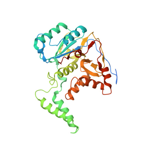
    The bacterial effector proteins SseK and NleB glycosylate host proteins on arginine residues, leading to reduced NF-κB-dependent responses to infection. Salmonella SseK1 and SseK2 are E. coli NleB1 orthologs that behave as NleB1-like GTs, although they differ in protein substrate specificity. Here we report that these enzymes are retaining glycosyltransferases composed of a helix-loop-helix (HLH) domain, a lid domain, and a catalytic domain. A conserved HEN motif (His-Glu-Asn) in the active site is important for enzyme catalysis and bacterial virulence. We observe differences between SseK1 and SseK2 in interactions with substrates and identify substrate residues that are critical for enzyme recognition. Long Molecular Dynamics simulations suggest that the HLH domain determines substrate specificity and the lid-domain regulates the opening of the active site. Overall, our data suggest a front-face S N i mechanism, explain differences in activities among these effectors, and have implications for future drug development against enteric pathogens.


  • Organizational Affiliation
    • Department of Systems Biology, College of Life Science and Biotechnology, Yonsei University, 50 Yonsei-ro, Seodaemun-gu, Seoul, 03722, Republic of Korea.

Macromolecule Content 

  • Total Structure Weight: 39.34 kDa 
  • Atom Count: 2,498 
  • Modeled Residue Count: 304 
  • Deposited Residue Count: 336 
  • Unique protein chains: 1

Macromolecules

Find similar proteins by:|  3D Structure
Entity ID: 1
MoleculeChains  Sequence LengthOrganismDetailsImage
Transferase336Escherichia coliMutation(s): 0 
EC: 2.4.1
UniProt
Find proteins for Q9L9J3 (Salmonella typhimurium (strain LT2 / SGSC1412 / ATCC 700720))
Explore Q9L9J3 
Go to UniProtKB:  Q9L9J3
Entity Groups
Sequence Clusters30% Identity50% Identity70% Identity90% Identity95% Identity100% Identity
UniProt GroupQ9L9J3
Sequence Annotations
Expand
Reference Sequence

Small Molecules

Experimental Data & Validation

Experimental Data

  • Method: X-RAY DIFFRACTION
  • Resolution: 3.64 Å
  • R-Value Free:  0.285 (Depositor), 0.284 (DCC) 
  • R-Value Work:  0.254 (Depositor), 0.255 (DCC) 
  • R-Value Observed: 0.255 (Depositor) 
Space Group: P 62 2 2
Unit Cell:
Length ( Å )Angle ( ˚ )
a = 115.94α = 90
b = 115.94β = 90
c = 100.08γ = 120
Software Package:
Software NamePurpose
REFMACrefinement
HKL-2000data reduction
HKL-2000data scaling
REFMACphasing

Structure Validation

View Full Validation Report


Ligand Structure Quality Assessment 


Entry History 

Deposition Data

Revision History  (Full details and data files)

  • Version 1.0: 2017-12-20
    Type: Initial release
  • Version 1.1: 2018-09-19
    Changes: Data collection, Database references
  • Version 1.2: 2018-10-24
    Changes: Data collection, Database references
  • Version 1.3: 2018-10-31
    Changes: Data collection, Database references
  • Version 1.4: 2024-11-06
    Changes: Data collection, Database references, Derived calculations, Structure summary