5H46 | pdb_00005h46

Mycobacterium smegmatis Dps1 mutant - F47E


Experimental Data Snapshot

  • Method: X-RAY DIFFRACTION
  • Resolution: 2.85 Å
  • R-Value Free: 
    0.260 (Depositor), 0.258 (DCC) 
  • R-Value Work: 
    0.229 (Depositor), 0.224 (DCC) 
  • R-Value Observed: 
    0.232 (Depositor) 

Starting Model: experimental
View more details

wwPDB Validation 3D Report Full Report

Validation slider image for 5H46

This is version 1.2 of the entry. See complete history

Literature

A Mutation Directs the Structural Switch of DNA Binding Proteins under Starvation to a Ferritin-like Protein Cage.

Williams, S.M.Chandran, A.V.Prakash, S.Vijayan, M.Chatterji, D.

(2017) Structure 25: 1449-1454.e3

  • DOI: https://doi.org/10.1016/j.str.2017.07.006
  • Primary Citation Related Structures: 
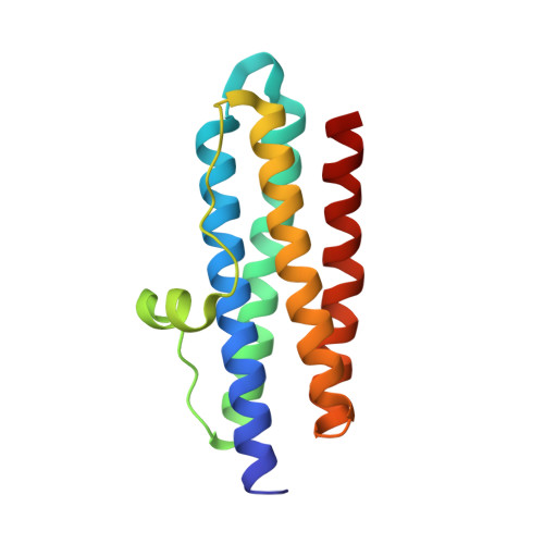
    5H46

  • PubMed Abstract: 

    Proteins of the ferritin family are ubiquitous in living organisms. With their spherical cage-like structures they are the iron storehouses in cells. Subfamilies of ferritins include 24-meric ferritins and bacterioferritins (maxiferritins), and 12-meric Dps (miniferritins). Dps safeguards DNA by direct binding, affording physical protection and safeguards from free radical-mediated damage by sequestering iron in its core. The maxiferritins can oxidize and store iron but cannot bind DNA. Here we show that a mutation at a critical interface in Dps alters its assembly from the canonical 12-mer to a ferritin-like 24-mer under crystallization. This structural switch was attributed to the conformational alteration of a highly conserved helical loop and rearrangement of the C-terminus. Our results demonstrate a novel concept of mutational switch between related protein subfamilies and corroborate the popular model for evolution by which subtle substitutions in an amino acid sequence lead to diversification among proteins.


  • Organizational Affiliation
    • Molecular Biophysics Unit, Indian Institute of Science, Bangalore 560 012, India.

Macromolecule Content 

  • Total Structure Weight: 16.98 kDa 
  • Atom Count: 1,185 
  • Modeled Residue Count: 149 
  • Deposited Residue Count: 149 
  • Unique protein chains: 1

Macromolecules

Find similar proteins by:|  3D Structure
Entity ID: 1
MoleculeChains  Sequence LengthOrganismDetailsImage
DNA protection during starvation protein149Mycolicibacterium smegmatis MC2 155Mutation(s): 1 
Gene Names: dpsMSMEG_6467MSMEI_6295
EC: 1.16
UniProt
Find proteins for A0R692 (Mycolicibacterium smegmatis (strain ATCC 700084 / mc(2)155))
Explore A0R692 
Go to UniProtKB:  A0R692
Entity Groups
Sequence Clusters30% Identity50% Identity70% Identity90% Identity95% Identity100% Identity
UniProt GroupA0R692
Sequence Annotations
Expand
Reference Sequence

Experimental Data & Validation

Experimental Data

  • Method: X-RAY DIFFRACTION
  • Resolution: 2.85 Å
  • R-Value Free:  0.260 (Depositor), 0.258 (DCC) 
  • R-Value Work:  0.229 (Depositor), 0.224 (DCC) 
  • R-Value Observed: 0.232 (Depositor) 
Space Group: F 4 3 2
Unit Cell:
Length ( Å )Angle ( ˚ )
a = 175.07α = 90
b = 175.07β = 90
c = 175.07γ = 90
Software Package:
Software NamePurpose
REFMACrefinement
REFMACdata reduction
REFMACdata scaling
Cootmodel building
REFMACphasing

Structure Validation

View Full Validation Report


Entry History 

& Funding Information

Deposition Data


Funding OrganizationLocationGrant Number
India--

Revision History  (Full details and data files)

  • Version 1.0: 2017-09-06
    Type: Initial release
  • Version 1.1: 2017-12-06
    Changes: Database references
  • Version 1.2: 2023-11-08
    Changes: Data collection, Database references, Refinement description