


Crystal structure of Human Dihydroorotate Dehydrogenase (DHODH) with 7GF
Huang, J., Wu, D.To be published.
Experimental Data Snapshot
Starting Model: experimental
View more details
Entity ID: 1 | |||||
|---|---|---|---|---|---|
| Molecule | Chains | Sequence Length | Organism | Details | Image |
| Dihydroorotate dehydrogenase (quinone), mitochondrial | 390 | Homo sapiens | Mutation(s): 0 Gene Names: DHODH EC: 1.3.5.2 | 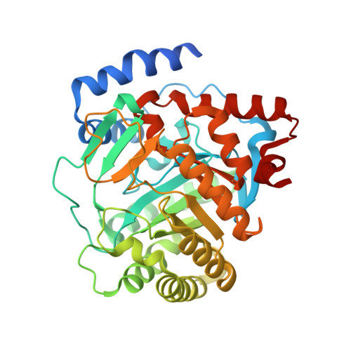 | |
UniProt & NIH Common Fund Data Resources | |||||
PHAROS: Q02127 GTEx: ENSG00000102967 | |||||
Entity Groups | |||||
| Sequence Clusters | 30% Identity50% Identity70% Identity90% Identity95% Identity100% Identity | ||||
| UniProt Group | Q02127 | ||||
Sequence AnnotationsExpand | |||||
Reference Sequence | |||||
| Ligands 5 Unique | |||||
|---|---|---|---|---|---|
| ID | Chains | Name / Formula / InChI Key | 2D Diagram | 3D Interactions | |
| FMN Download:Ideal Coordinates CCD File | B [auth A] | FLAVIN MONONUCLEOTIDE C17 H21 N4 O9 P FVTCRASFADXXNN-SCRDCRAPSA-N |  | ||
| 7GF Download:Ideal Coordinates CCD File | H [auth A] | methyl (2Z)-3-azanyl-2-[3-(4-bromophenyl)-4-oxidanylidene-1,3-thiazolidin-2-ylidene]propanoate C13 H13 Br N2 O3 S UEDYOLMWFOHHRV-BENRWUELSA-N |  | ||
| ORO Download:Ideal Coordinates CCD File | C [auth A] | OROTIC ACID C5 H4 N2 O4 PXQPEWDEAKTCGB-UHFFFAOYSA-N |  | ||
| SO4 Download:Ideal Coordinates CCD File | D [auth A], E [auth A] | SULFATE ION O4 S QAOWNCQODCNURD-UHFFFAOYSA-L |  | ||
| ACT Download:Ideal Coordinates CCD File | F [auth A], G [auth A] | ACETATE ION C2 H3 O2 QTBSBXVTEAMEQO-UHFFFAOYSA-M |  | ||
| Length ( Å ) | Angle ( ˚ ) |
|---|---|
| a = 90.875 | α = 90 |
| b = 90.875 | β = 90 |
| c = 123.17 | γ = 120 |
| Software Name | Purpose |
|---|---|
| REFMAC | refinement |
| CrysalisPro | data processing |
| HKL-3000 | data scaling |
| PHENIX | data collection |


