5H2Y | pdb_00005h2y

Crystal structure of reduced DapF from Corynebacterium glutamicum


Experimental Data Snapshot

  • Method: X-RAY DIFFRACTION
  • Resolution: 2.00 Å
  • R-Value Free: 
    0.252 (Depositor), 0.260 (DCC) 
  • R-Value Work: 
    0.207 (Depositor), 0.221 (DCC) 
  • R-Value Observed: 
    0.209 (Depositor) 

wwPDB Validation 3D Report Full Report

Validation slider image for 5H2Y

This is version 1.3 of the entry. See complete history

Literature

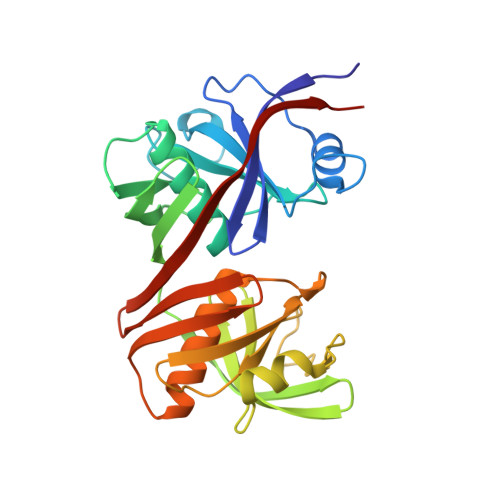
Structural basis for redox sensitivity in Corynebacterium glutamicum diaminopimelate epimerase: an enzyme involved in l-lysine biosynthesis.

Sagong, H.Y.Kim, K.J.

(2017) Sci Rep 7: 42318-42318

  • DOI: https://doi.org/10.1038/srep42318
  • Primary Citation Related Structures: 
    5H2G, 5H2Y, 5M47

  • PubMed Abstract: 

    Diaminopimelate epimerase (DapF) is one of the crucial enzymes involved in l-lysine biosynthesis, where it converts l,l-diaminopimelate (l,l-DAP) into d,l-DAP. DapF is also considered as an attractive target for the development of antibacterial drugs. Here, we report the crystal structure of DapF from Corynebacterium glutamicum (CgDapF). Structures of CgDapF obtained under both oxidized and reduced conditions reveal that the function of CgDapF is regulated by redox-switch modulation via reversible disulfide bond formation between two catalytic cysteine residues. Under oxidized condition, two catalytic cysteine residues form a disulfide bond; these same cysteine residues exist in reduced form under reduced condition. Disulfide bond formation also induces a subsequent structural change in the dynamic catalytic loop at the active site, which results in open/closed conformational change at the active site. We also determined the crystal structure of CgDapF in complex with its product d,l-DAP, and elucidated how the enzyme recognizes its substrate l,l-DAP as a substrate. Moreover, the structure in complex with the d,l-DAP product reveals that CgDapF undergoes a large open/closed domain movement upon substrate binding, resulting in a completely buried active site with the substrate bound.


  • Organizational Affiliation
    • School of Life Sciences, KNU Creative BioResearch Group, Kyungpook National University, Daehak-ro 80, Buk-ku, Daegu 702-701, Korea.

Macromolecule Content 

  • Total Structure Weight: 60.11 kDa 
  • Atom Count: 4,273 
  • Modeled Residue Count: 554 
  • Deposited Residue Count: 566 
  • Unique protein chains: 1

Macromolecules

Find similar proteins by:|  3D Structure
Entity ID: 1
MoleculeChains  Sequence LengthOrganismDetailsImage
Diaminopimelate epimerase
A, B
283Corynebacterium glutamicum ATCC 13032Mutation(s): 0 
Gene Names: dapFCgl1943cg2129
EC: 5.1.1.7
UniProt
Find proteins for Q8NP73 (Corynebacterium glutamicum (strain ATCC 13032 / DSM 20300 / JCM 1318 / BCRC 11384 / CCUG 27702 / LMG 3730 / NBRC 12168 / NCIMB 10025 / NRRL B-2784 / 534))
Explore Q8NP73 
Go to UniProtKB:  Q8NP73
Entity Groups
Sequence Clusters30% Identity50% Identity70% Identity90% Identity95% Identity100% Identity
UniProt GroupQ8NP73
Sequence Annotations
Expand
Reference Sequence

Experimental Data & Validation

Experimental Data

  • Method: X-RAY DIFFRACTION
  • Resolution: 2.00 Å
  • R-Value Free:  0.252 (Depositor), 0.260 (DCC) 
  • R-Value Work:  0.207 (Depositor), 0.221 (DCC) 
  • R-Value Observed: 0.209 (Depositor) 
Space Group: I 2 2 2
Unit Cell:
Length ( Å )Angle ( ˚ )
a = 101.757α = 90
b = 118.938β = 90
c = 153.333γ = 90
Software Package:
Software NamePurpose
REFMACrefinement
HKL-2000data collection
PDB_EXTRACTdata extraction
HKL-2000data reduction
HKL-2000data scaling
PHENIXphasing
HKLdata reduction

Structure Validation

View Full Validation Report


Entry History 

Deposition Data

Revision History  (Full details and data files)

  • Version 1.0: 2016-11-09
    Type: Initial release
  • Version 1.1: 2017-07-19
    Changes: Database references, Derived calculations
  • Version 1.2: 2020-09-09
    Changes: Structure summary
  • Version 1.3: 2024-03-20
    Changes: Data collection, Database references