5H0G

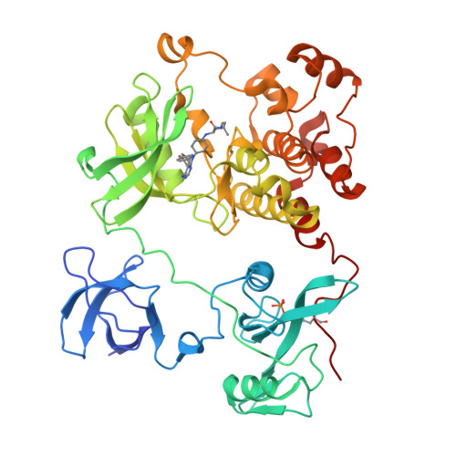
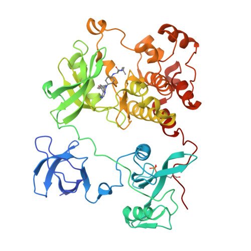
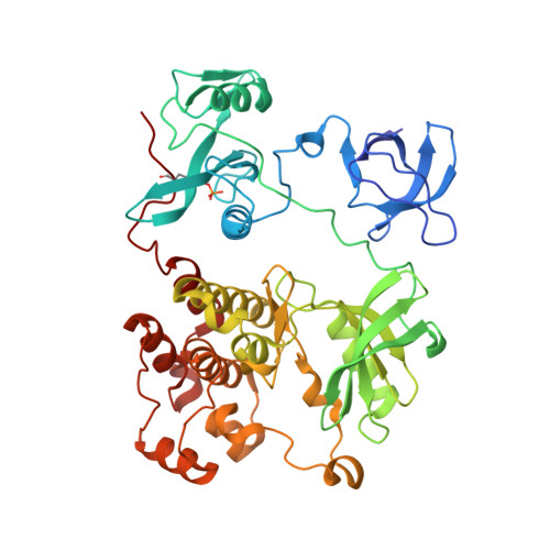
Crystal structure of HCK complexed with a pyrrolo-pyrimidine inhibitor (S)-2-(((1r,4S)-4-(4-amino-5-(4-phenoxyphenyl)-7H-pyrrolo[2,3-d]pyrimidin-7-yl)cyclohexyl)amino)-N,4-dimethylpentanamide


Experimental Data Snapshot

  • Method: X-RAY DIFFRACTION
  • Resolution: 1.80 Å
  • R-Value Free: 0.224 
  • R-Value Work: 0.181 
  • R-Value Observed: 0.183 

Starting Model: experimental
View more details

wwPDB Validation   3D Report Full Report


Ligand Structure Quality Assessment 


This is version 1.2 of the entry. See complete history


Literature

Activity cliff for 7-substituted pyrrolo-pyrimidine inhibitors of HCK explained in terms of predicted basicity of the amine nitrogen.

Yuki, H.Kikuzato, K.Koda, Y.Mikuni, J.Tomabechi, Y.Kukimoto-Niino, M.Tanaka, A.Shirai, F.Shirouzu, M.Koyama, H.Honma, T.

(2017) Bioorg Med Chem 25: 4259-4264

  • DOI: https://doi.org/10.1016/j.bmc.2017.05.053
  • Primary Citation of Related Structures:  
    5H09, 5H0B, 5H0E, 5H0G, 5H0H

  • PubMed Abstract: 

    We previously reported the structure-based design of a highly potent hematopoietic cell kinase (HCK) inhibitor, a pyrrolo-pyrimidine compound designated RK-20449, for treatment of recurrent leukemia. Herein we report the synthesis and structure-activity relationships of some amino acid derivatives of 7-substituted pyrrolo-pyrimidine. Although these derivatives had the same predicted binding conformation as RK-20449, their IC 50 values were 100-1000 times larger than that of the parent compound. We assumed that the basicity of the amine nitrogen, which formed an ionic bond with Asp348 of HCK, markedly affected inhibitory activity against HCK. The pKa values of the nitrogen were predicted by means of an ab initio quantum mechanical method, and complexes of the derivatives with HCK were analyzed by X-ray crystallography. We observed a significant correlation between the predicted pKa and IC 50 values, and the crystal structures of the less potent derivatives showed various types of defects around the ionic bond.


  • Organizational Affiliation

    Drug Discovery Computational Chemistry Platform Unit, RIKEN Center for Life Science Technologies, 1-7-22 Suehiro-cho, Tsurumi-ku, Yokohama, Kanagawa 230-0045, Japan.


Macromolecules
Find similar proteins by:  (by identity cutoff)  |  3D Structure
Entity ID: 1
MoleculeChains Sequence LengthOrganismDetailsImage
Tyrosine-protein kinase HCK454Homo sapiensMutation(s): 3 
Gene Names: HCK
EC: 2.7.10.2
UniProt & NIH Common Fund Data Resources
Find proteins for P08631 (Homo sapiens)
Go to UniProtKB:  P08631
PHAROS:  P08631
GTEx:  ENSG00000101336 
Sequence Annotations
Expand
  • Reference Sequence
Small Molecules
Ligands 1 Unique
IDChains Name / Formula / InChI Key2D Diagram3D Interactions
OOU
Query on OOU

Download Ideal Coordinates CCD File 
B [auth A](2~{S})-2-[[4-[4-azanyl-5-(4-phenoxyphenyl)pyrrolo[2,3-d]pyrimidin-7-yl]cyclohexyl]amino]-~{N},4-dimethyl-pentanamide
C31 H38 N6 O2
QWSTVNCCNITNGB-WCYRKSIYSA-N
Modified Residues  1 Unique
IDChains TypeFormula2D DiagramParent
PTR
Query on PTR
A
L-PEPTIDE LINKINGC9 H12 N O6 PTYR
Binding Affinity Annotations 
IDSourceBinding Affinity
OOU BindingDB:  5H0G IC50: min: 111, max: 112 (nM) from 2 assay(s)
Experimental Data & Validation

Experimental Data

  • Method: X-RAY DIFFRACTION
  • Resolution: 1.80 Å
  • R-Value Free: 0.224 
  • R-Value Work: 0.181 
  • R-Value Observed: 0.183 
  • Space Group: P 21 21 21
Unit Cell:
Length ( Å )Angle ( ˚ )
a = 43.072α = 90
b = 85.338β = 90
c = 129.717γ = 90
Software Package:
Software NamePurpose
DENZOdata collection
HKL-2000data scaling
PHASERphasing
PHENIXrefinement
PDB_EXTRACTdata extraction
HKL-2000data reduction

Structure Validation

View Full Validation Report


Ligand Structure Quality Assessment 


Entry History 

Deposition Data

Revision History  (Full details and data files)

  • Version 1.0: 2017-10-04
    Type: Initial release
  • Version 1.1: 2023-11-08
    Changes: Data collection, Database references, Refinement description
  • Version 1.2: 2023-11-15
    Changes: Data collection