


Acryloyl-CoA reductase AcuI from Ruegeria pomeroyi DSS-3
Zhang, Y.Z., Wang, P., Cao, H.Y.To be published.
Experimental Data Snapshot
wwPDB Validation 3D Report Full Report

Entity ID: 1 | |||||
|---|---|---|---|---|---|
| Molecule | Chains | Sequence Length | Organism | Details | Image |
| Acrylyl-CoA reductase AcuI | 330 | Ruegeria pomeroyi DSS-3 | Mutation(s): 0 Gene Names: acuI, SPO1914 EC: 1.3.1.84 | 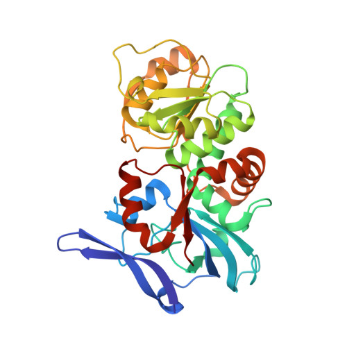 | |
UniProt | |||||
Entity Groups | |||||
| Sequence Clusters | 30% Identity50% Identity70% Identity90% Identity95% Identity100% Identity | ||||
| UniProt Group | Q5LS56 | ||||
Sequence AnnotationsExpand | |||||
Reference Sequence | |||||
| Ligands 1 Unique | |||||
|---|---|---|---|---|---|
| ID | Chains | Name / Formula / InChI Key | 2D Diagram | 3D Interactions | |
| BR Download:Ideal Coordinates CCD File | C [auth A], D [auth B] | BROMIDE ION Br CPELXLSAUQHCOX-UHFFFAOYSA-M |  | ||
| Length ( Å ) | Angle ( ˚ ) |
|---|---|
| a = 91.841 | α = 90 |
| b = 111.218 | β = 90 |
| c = 75.241 | γ = 90 |
| Software Name | Purpose |
|---|---|
| PHENIX | refinement |
| HKL-2000 | data reduction |
| HKL-2000 | data scaling |
| PHASER | phasing |


