5GQH | pdb_00005gqh

Cryo-EM structure of PaeCas3-AcrF3 complex


Experimental Data Snapshot

  • Method: ELECTRON MICROSCOPY
  • Resolution: 4.20 Å
  • Aggregation State: PARTICLE 
  • Reconstruction Method: SINGLE PARTICLE 

wwPDB Validation 3D Report Full Report

Validation slider image for 5GQH

This is version 1.3 of the entry. See complete history

Literature

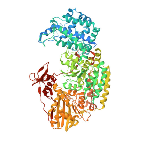
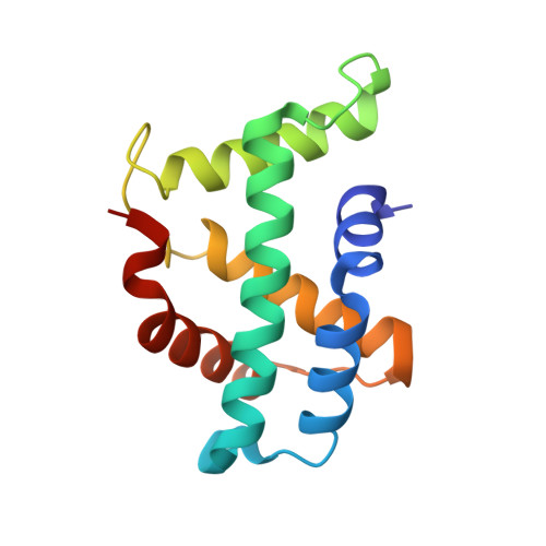
A CRISPR evolutionary arms race: structural insights into viral anti-CRISPR/Cas responses

Wang, J.Ma, J.Cheng, Z.Meng, X.You, L.Wang, M.Zhang, X.Wang, Y.

(2016) Cell Res 26: 1165-1168

  • DOI: https://doi.org/10.1038/cr.2016.103
  • Primary Citation Related Structures: 
    5GNF, 5GQH

  • Organizational Affiliation
    • Key Laboratory of RNA Biology, Beijing 100101, China.

Macromolecule Content 

  • Total Structure Weight: 154.51 kDa 
  • Atom Count: 6,641 
  • Modeled Residue Count: 1,171 
  • Deposited Residue Count: 1,368 
  • Unique protein chains: 2

Macromolecules

Find similar proteins by:|  3D Structure
Entity ID: 1
MoleculeChains  Sequence LengthOrganismDetailsImage
CRISPR-associated nuclease/helicase Cas3 subtype I-F/YPEST1,090Pseudomonas aeruginosa UCBPP-PA14Mutation(s): 0 
Gene Names: cas3
EC: 3.1 (PDB Primary Data), 3.6.4 (PDB Primary Data)
UniProt
Find proteins for Q02ML8 (Pseudomonas aeruginosa (strain UCBPP-PA14))
Explore Q02ML8 
Go to UniProtKB:  Q02ML8
Entity Groups
Sequence Clusters30% Identity50% Identity70% Identity90% Identity95% Identity100% Identity
UniProt GroupQ02ML8
Sequence Annotations
Expand
Reference Sequence
Find similar proteins by:|  3D Structure
Entity ID: 2
MoleculeChains  Sequence LengthOrganismDetailsImage
anti-CRISPR protein 3
B, C
139Pseudomonas phage JBD5Mutation(s): 0 
Gene Names: JBD5_035
UniProt
Find proteins for L7P7R7 (Pseudomonas phage JBD5)
Explore L7P7R7 
Go to UniProtKB:  L7P7R7
Entity Groups
Sequence Clusters30% Identity50% Identity70% Identity90% Identity95% Identity100% Identity
UniProt GroupL7P7R7
Sequence Annotations
Expand
Reference Sequence

Experimental Data & Validation

Experimental Data

  • Method: ELECTRON MICROSCOPY
  • Resolution: 4.20 Å
  • Aggregation State: PARTICLE 
  • Reconstruction Method: SINGLE PARTICLE 

Structure Validation

View Full Validation Report


Entry History 

Deposition Data

Revision History  (Full details and data files)

  • Version 1.0: 2016-09-21
    Type: Initial release
  • Version 1.1: 2016-10-19
    Changes: Database references
  • Version 1.2: 2019-11-06
    Changes: Data collection, Other
  • Version 1.3: 2024-03-20
    Changes: Data collection, Database references