5GMG | pdb_00005gmg

Crystal structure of monkey TLR7 in complex with loxoribine and polyU


Experimental Data Snapshot

  • Method: X-RAY DIFFRACTION
  • Resolution: 2.60 Å
  • R-Value Free: 
    0.258 (Depositor), 0.261 (DCC) 
  • R-Value Work: 
    0.215 (Depositor), 0.216 (DCC) 
  • R-Value Observed: 
    0.217 (Depositor) 

Starting Model: experimental
View more details

wwPDB Validation 3D Report Full Report

Validation slider image for 5GMG

Ligand Structure Quality Assessment 


This is version 2.2 of the entry. See complete history

Literature

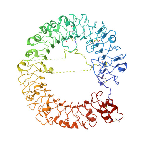
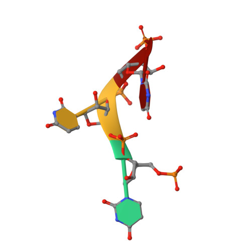
Structural Analysis Reveals that Toll-like Receptor 7 Is a Dual Receptor for Guanosine and Single-Stranded RNA

Zhang, Z.Ohto, U.Shibata, T.Krayukhina, E.Taoka, M.Yamauchi, Y.Tanji, H.Isobe, T.Uchiyama, S.Miyake, K.Shimizu, T.

(2016) Immunity 45: 737-748

  • DOI: https://doi.org/10.1016/j.immuni.2016.09.011
  • Primary Citation Related Structures: 
    5GMF, 5GMG, 5GMH

  • PubMed Abstract: 

    Toll-like receptor 7 (TLR7) is a single-stranded RNA (ssRNA) sensor in innate immunity and also responds to guanosine and chemical ligands, such as imidazoquinoline compounds. However, TLR7 activation mechanism by these ligands remain largely unknown. Here, we generated crystal structures of three TLR7 complexes, and found that all formed an activated m-shaped dimer with two ligand-binding sites. The first site conserved in TLR7 and TLR8 was used for small ligand-binding essential for its activation. The second site spatially distinct from that of TLR8 was used for a ssRNA-binding that enhanced the affinity of the first-site ligands. The first site preferentially recognized guanosine and the second site specifically bound to uridine moieties in ssRNA. Our structural, biochemical, and mutagenesis studies indicated that TLR7 is a dual receptor for guanosine and uridine-containing ssRNA. Our findings have important implications for understanding of TLR7 function, as well as for therapeutic manipulation of TLR7 activation.


  • Organizational Affiliation
    • Graduate School of Pharmaceutical Sciences, The University of Tokyo, 7-3-1 Hongo, Bunkyo-ku, Tokyo 113-0033, Japan.

Macromolecule Content 

  • Total Structure Weight: 193.34 kDa 
  • Atom Count: 12,680 
  • Modeled Residue Count: 1,532 
  • Deposited Residue Count: 1,642 
  • Unique protein chains: 1
  • Unique nucleic acid chains: 1

Macromolecules


Find similar proteins by:|  3D Structure
Entity ID: 1
MoleculeChains  Sequence LengthOrganismDetailsImage
Toll-like receptor 7
A, B
817Macaca mulattaMutation(s): 4 
Gene Names: TLR7
UniProt
Find proteins for B3Y653 (Macaca mulatta)
Explore B3Y653 
Go to UniProtKB:  B3Y653
Entity Groups
Sequence Clusters30% Identity50% Identity70% Identity90% Identity95% Identity100% Identity
UniProt GroupB3Y653
Glycosylation
Glycosylation Sites: 4
Sequence Annotations
Expand
Reference Sequence
Find similar nucleic acids by:  Sequence
Entity ID: 2
MoleculeChains LengthOrganismImage
RNA (5'-R(P*UP*UP*UP*U)-3')
C, D
4Homo sapiens
Sequence Annotations
Expand
Reference Sequence

Oligosaccharides

Help  
Entity ID: 3
MoleculeChains Length2D Diagram GlycosylationD Interactions
2-acetamido-2-deoxy-beta-D-glucopyranose-(1-4)-2-acetamido-2-deoxy-beta-D-glucopyranose
E, F
2N-Glycosylation
Glycosylation Resources
GlyTouCan: G42666HT
GlyCosmos: G42666HT
GlyGen: G42666HT

Small Molecules

Ligands 3 Unique
IDChains Name / Formula / InChI Key2D Diagram3D Interactions
SDL

Query on SDL


Download:Ideal Coordinates CCD File
J [auth B],
K [auth B]
2-azanyl-9-[(2~{R},3~{R},4~{S},5~{R})-5-(hydroxymethyl)-3,4-bis(oxidanyl)oxolan-2-yl]-7-prop-2-enyl-1~{H}-purine-6,8-dione
C13 H17 N5 O6
VDCRFBBZFHHYGT-IOSLPCCCSA-N
NAG

Query on NAG


Download:Ideal Coordinates CCD File
G [auth A]
H [auth A]
I [auth A]
L [auth B]
M [auth B]
G [auth A],
H [auth A],
I [auth A],
L [auth B],
M [auth B],
N [auth B]
2-acetamido-2-deoxy-beta-D-glucopyranose
C8 H15 N O6
OVRNDRQMDRJTHS-FMDGEEDCSA-N
PO4

Query on PO4


Download:Ideal Coordinates CCD File
O [auth B]PHOSPHATE ION
O4 P
NBIIXXVUZAFLBC-UHFFFAOYSA-K
Binding Affinity Annotations 
IDSourceBinding Affinity
SDL BindingDB:  5GMG EC50: 3.22e+5 (nM) from 1 assay(s)

Experimental Data & Validation

Experimental Data

  • Method: X-RAY DIFFRACTION
  • Resolution: 2.60 Å
  • R-Value Free:  0.258 (Depositor), 0.261 (DCC) 
  • R-Value Work:  0.215 (Depositor), 0.216 (DCC) 
  • R-Value Observed: 0.217 (Depositor) 
Space Group: P 1 21 1
Unit Cell:
Length ( Å )Angle ( ˚ )
a = 98.277α = 90
b = 99.41β = 98.58
c = 112.096γ = 90
Software Package:
Software NamePurpose
REFMACrefinement
XDSdata reduction
XDSdata scaling
MOLREPphasing

Structure Validation

View Full Validation Report


Ligand Structure Quality Assessment 


Entry History 

Deposition Data

Revision History  (Full details and data files)

  • Version 1.0: 2016-11-02
    Type: Initial release
  • Version 1.1: 2020-02-26
    Changes: Data collection, Database references, Derived calculations
  • Version 2.0: 2020-07-29
    Type: Remediation
    Reason: Carbohydrate remediation
    Changes: Atomic model, Data collection, Derived calculations, Structure summary
  • Version 2.1: 2023-11-08
    Changes: Data collection, Database references, Refinement description, Structure summary
  • Version 2.2: 2024-10-23
    Changes: Structure summary