5GKR | pdb_00005gkr

Crystal structure of SLE patient-derived anti-DNA antibody in complex with oligonucleotide


Experimental Data Snapshot

  • Method: X-RAY DIFFRACTION
  • Resolution: 2.10 Å
  • R-Value Free: 
    0.185 (Depositor), 0.179 (DCC) 
  • R-Value Work: 
    0.163 (Depositor) 
  • R-Value Observed: 
    0.164 (Depositor) 

Starting Models: experimental
View more details

wwPDB Validation 3D Report Full Report

Validation slider image for 5GKR

This is version 1.3 of the entry. See complete history

Literature

Clonal evolution and antigen recognition of anti-nuclear antibodies in acute systemic lupus erythematosus

Sakakibara, S.Arimori, T.Yamashita, K.Jinzai, H.Motooka, D.Nakamura, S.Li, S.Takeda, K.Katayama, J.El Hussien, M.A.Narazaki, M.Tanaka, T.Standley, D.M.Takagi, J.Kikutani, H.

(2017) Sci Rep 7: 16428-16428

  • DOI: https://doi.org/10.1038/s41598-017-16681-y
  • Primary Citation Related Structures: 
    5GKR, 5GKS

  • PubMed Abstract: 

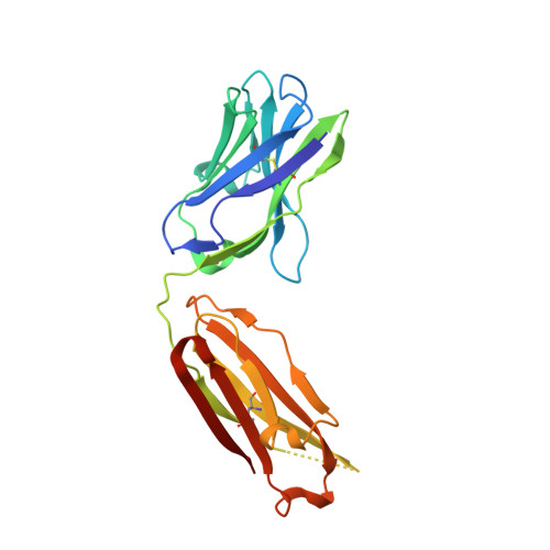
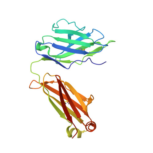
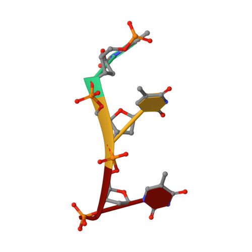
    The evolutional process of disease-associated autoantibodies in systemic lupus erythematosus (SLE) remains to be established. Here we show intraclonal diversification and affinity maturation of anti-nuclear antibody (ANA)-producing B cells in SLE. We identified a panel of monoclonal ANAs recognizing nuclear antigens, such as double-stranded DNA (dsDNA) and ribonucleoproteins (RNPs) from acute SLE subjects. These ANAs had relatively few, but nonetheless critical mutations. High-throughput immunoglobulin sequencing of blood lymphocytes disclosed the existence of sizable ANA lineages shearing critical mutations intraclonally. We further focused on anti-DNA antibodies, which are capable to bind to both single-stranded (ss) and dsDNA at high affinity. Crystal structure and biochemical analysis confirmed a direct role of the mutations in the acquisition of DNA reactivity and also revealed that these anti-DNA antibodies recognized an unpaired region within DNA duplex. Our study unveils the unique properties of high-affinity anti-DNA antibodies that are generated through antigen-driven affinity maturation in acute phase of SLE.


  • Organizational Affiliation
    • Laboratory of Immune Regulation, Immunology Frontier Research Center, Osaka University, Suita, Osaka, 565-0871, Japan. sakakibara@ifrec.osaka-u.ac.jp.

Macromolecule Content 

  • Total Structure Weight: 93.86 kDa 
  • Atom Count: 6,459 
  • Modeled Residue Count: 832 
  • Deposited Residue Count: 872 
  • Unique protein chains: 2
  • Unique nucleic acid chains: 1

Macromolecules


Find similar proteins by:|  3D Structure
Entity ID: 1
MoleculeChains  Sequence LengthOrganismDetailsImage
IgG2, Fab (heavy chain)
A, C
218Homo sapiensMutation(s): 0 
Find similar proteins by:|  3D Structure
Entity ID: 2
MoleculeChains  Sequence LengthOrganismDetailsImage
lambda, Fab (light chain)
B, D
216Homo sapiensMutation(s): 0 
Find similar nucleic acids by:  Sequence
Entity ID: 3
MoleculeChains LengthOrganismImage
DNA (5'-D(P*TP*TP*TP*T)-3')E [auth T]4Homo sapiens
Sequence Annotations
Expand
Reference Sequence

Experimental Data & Validation

Experimental Data

  • Method: X-RAY DIFFRACTION
  • Resolution: 2.10 Å
  • R-Value Free:  0.185 (Depositor), 0.179 (DCC) 
  • R-Value Work:  0.163 (Depositor) 
  • R-Value Observed: 0.164 (Depositor) 
Space Group: H 3
Unit Cell:
Length ( Å )Angle ( ˚ )
a = 147.515α = 90
b = 147.515β = 90
c = 112.807γ = 120
Software Package:
Software NamePurpose
REFMACrefinement
HKL-2000data reduction
HKL-2000data scaling
PHASERphasing

Structure Validation

View Full Validation Report


Entry History 

Deposition Data

Revision History  (Full details and data files)

  • Version 1.0: 2017-07-05
    Type: Initial release
  • Version 1.1: 2017-12-06
    Changes: Database references
  • Version 1.2: 2023-11-08
    Changes: Data collection, Database references, Refinement description
  • Version 1.3: 2024-10-23
    Changes: Structure summary