5GIS | pdb_00005gis

Crystal structure of a Fab fragment with its ligand peptide


Experimental Data Snapshot

  • Method: X-RAY DIFFRACTION
  • Resolution: 1.93 Å
  • R-Value Free: 
    0.216 (Depositor), 0.216 (DCC) 
  • R-Value Work: 
    0.186 (Depositor), 0.186 (DCC) 
  • R-Value Observed: 
    0.188 (Depositor) 

Starting Model: experimental
View more details

wwPDB Validation 3D Report Full Report

Validation slider image for 5GIS

This is version 2.1 of the entry. See complete history

Literature

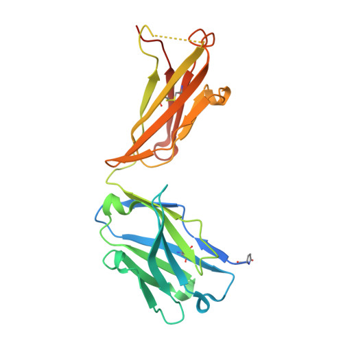
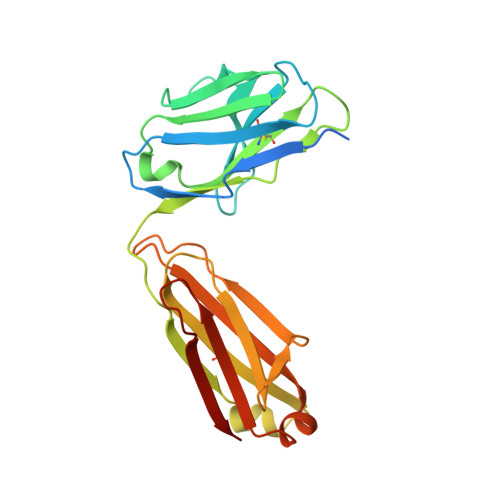
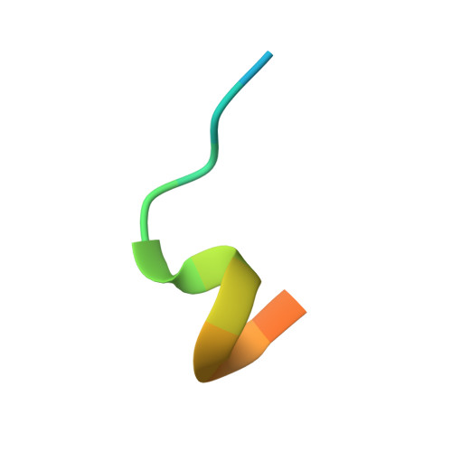
Structural basis for multi-specific peptide recognition by the anti-IDH1/2 monoclonal antibody, MsMab-1.

Kitago, Y.Kaneko, M.K.Ogasawara, S.Kato, Y.Takagi, J.

(2016) Biochem Biophys Res Commun 478: 1274-1279

  • DOI: https://doi.org/10.1016/j.bbrc.2016.08.110
  • Primary Citation Related Structures: 
    5GIR, 5GIS

  • PubMed Abstract: 

    A point mutation in isocitrate dehydrogenase 1 (IDH1) and IDH2 is directly linked to the pathogenesis of certain types of tumors. To detect this mutation, several antibodies that can distinguish between mutant and wild-type enzymes have been established. One of which, MsMab-1, has a unique multi-specific character against several types of mutated IDH1/2. This promiscuous character is in remarkable contrast to the highly specific antigen recognition typically observed with a monoclonal antibody. We solved the crystal structure of MsMab-1 Fab fragment in complex with either IDH1 or IDH2-derived peptides. Based on the structure, it became clear that the peptide-binding pocket of the antibody is highly complementary to the core determinant shared between the IDH1 and IDH2, while leaving just enough space for the side chain of the pathogenic but not the wild-type amino acids located in the mutation position. Clarification of the molecular basis for the peculiar binding characteristics of MsMab-1 in atomic detail will help facilitating its diagnostic application, and may be used to develop better diagnostic reagents through structure-guided protein engineering.


  • Organizational Affiliation
    • Laboratory of Protein Synthesis and Expression, Institute for Protein Research, Osaka University, 3-2 Yamadaoka, Suita, Osaka 565-0871, Japan.

Macromolecule Content 

  • Total Structure Weight: 53.76 kDa 
  • Atom Count: 3,591 
  • Modeled Residue Count: 432 
  • Deposited Residue Count: 490 
  • Unique protein chains: 3

Macromolecules

Find similar proteins by:|  3D Structure
Entity ID: 1
MoleculeChains  Sequence LengthOrganismDetailsImage
Heavy chain of Fab fragmentA [auth H]240Mus musculusMutation(s): 0 
Find similar proteins by:|  3D Structure
Entity ID: 2
MoleculeChains  Sequence LengthOrganismDetailsImage
Light chain of Fab fragmentB [auth L]234Mus musculusMutation(s): 0 
Find similar proteins by:  Sequence   |   3D Structure  
Entity ID: 3
MoleculeChains  Sequence LengthOrganismDetailsImage
THR-LYS-PRO-ILE-THR-ILE-GLY-SER-HIS-ALA-HIS-GLY-ASP-GLN-TYR-LYS16Homo sapiensMutation(s): 0 
EC: 1.1.1.42
UniProt & NIH Common Fund Data Resources
Find proteins for P48735 (Homo sapiens)
Explore P48735 
Go to UniProtKB:  P48735
PHAROS:  P48735
GTEx:  ENSG00000182054 
Entity Groups
Sequence Clusters30% Identity50% Identity70% Identity90% Identity95% Identity100% Identity
UniProt GroupP48735
Sequence Annotations
Expand
Reference Sequence

Experimental Data & Validation

Experimental Data

  • Method: X-RAY DIFFRACTION
  • Resolution: 1.93 Å
  • R-Value Free:  0.216 (Depositor), 0.216 (DCC) 
  • R-Value Work:  0.186 (Depositor), 0.186 (DCC) 
  • R-Value Observed: 0.188 (Depositor) 
Space Group: P 1 21 1
Unit Cell:
Length ( Å )Angle ( ˚ )
a = 57.05α = 90
b = 65β = 111.8
c = 67.13γ = 90
Software Package:
Software NamePurpose
REFMACrefinement
XDSdata processing
XDSdata scaling
PHASERphasing

Structure Validation

View Full Validation Report


Entry History 

Deposition Data

Revision History  (Full details and data files)

  • Version 1.0: 2016-09-14
    Type: Initial release
  • Version 1.1: 2017-12-06
    Changes: Data collection, Database references, Derived calculations
  • Version 2.0: 2023-11-08
    Changes: Data collection, Database references, Polymer sequence, Refinement description
  • Version 2.1: 2024-11-13
    Changes: Structure summary