5GGI | pdb_00005ggi

Crystal structure of human protein O-mannose beta-1,2-N-acetylglucosaminyltransferase in complex with Mn, UDP and Mannosyl-peptide


Experimental Data Snapshot

  • Method: X-RAY DIFFRACTION
  • Resolution: 2.60 Å
  • R-Value Free: 
    0.238 (Depositor), 0.244 (DCC) 
  • R-Value Work: 
    0.199 (Depositor), 0.207 (DCC) 
  • R-Value Observed: 
    0.201 (Depositor) 

Starting Model: experimental
View more details

wwPDB Validation 3D Report Full Report

Validation slider image for 5GGI

Ligand Structure Quality Assessment 


This is version 1.6 of the entry. See complete history

Literature

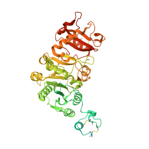
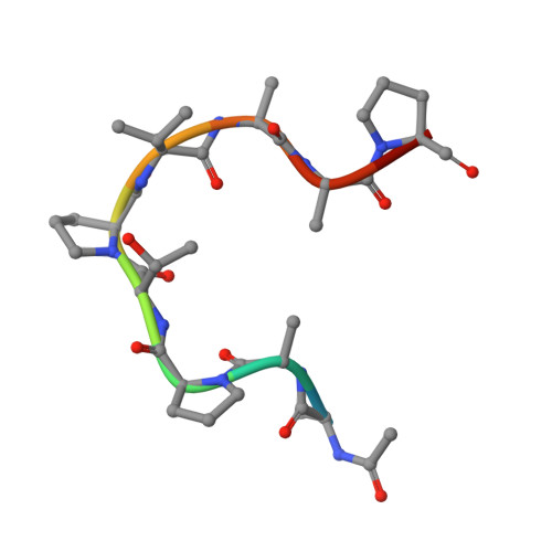
Carbohydrate-binding domain of the POMGnT1 stem region modulates O-mannosylation sites of alpha-dystroglycan

Kuwabara, N.Manya, H.Yamada, T.Tateno, H.Kanagawa, M.Kobayashi, K.Akasaka-Manya, K.Hirose, Y.Mizuno, M.Ikeguchi, M.Toda, T.Hirabayashi, J.Senda, T.Endo, T.Kato, R.

(2016) Proc Natl Acad Sci U S A 113: 9280-9285

  • DOI: https://doi.org/10.1073/pnas.1525545113
  • Primary Citation Related Structures: 
    5GGF, 5GGG, 5GGI, 5GGJ, 5GGK, 5GGL, 5GGN, 5GGO, 5GGP

  • PubMed Abstract: 

    The dystrophin glycoprotein complex, which connects the cell membrane to the basement membrane, is essential for a variety of biological events, including maintenance of muscle integrity. An O-mannose-type GalNAc-β1,3-GlcNAc-β1,4-(phosphate-6)-Man structure of α-dystroglycan (α-DG), a subunit of the complex that is anchored to the cell membrane, interacts directly with laminin in the basement membrane. Reduced glycosylation of α-DG is linked to some types of inherited muscular dystrophy; consistent with this relationship, many disease-related mutations have been detected in genes involved in O-mannosyl glycan synthesis. Defects in protein O-linked mannose β1,2-N-acetylglucosaminyltransferase 1 (POMGnT1), a glycosyltransferase that participates in the formation of GlcNAc-β1,2-Man glycan, are causally related to muscle-eye-brain disease (MEB), a congenital muscular dystrophy, although the role of POMGnT1 in postphosphoryl modification of GalNAc-β1,3-GlcNAc-β1,4-(phosphate-6)-Man glycan remains elusive. Our crystal structures of POMGnT1 agreed with our previous results showing that the catalytic domain recognizes substrate O-mannosylated proteins via hydrophobic interactions with little sequence specificity. Unexpectedly, we found that the stem domain recognizes the β-linked GlcNAc of O-mannosyl glycan, an enzymatic product of POMGnT1. This interaction may recruit POMGnT1 to a specific site of α-DG to promote GlcNAc-β1,2-Man clustering and also may recruit other enzymes that interact with POMGnT1, e.g., fukutin, which is required for further modification of the GalNAc-β1,3-GlcNAc-β1,4-(phosphate-6)-Man glycan. On the basis of our findings, we propose a mechanism for the deficiency in postphosphoryl modification of the glycan observed in POMGnT1-KO mice and MEB patients.


  • Organizational Affiliation
    • Photon Factory, Structural Biology Research Center, Institute of Materials Structure Science, High Energy Accelerator Research Organization, Tsukuba, Ibaraki 305-0801, Japan;

Macromolecule Content 

  • Total Structure Weight: 134.11 kDa 
  • Atom Count: 7,946 
  • Modeled Residue Count: 960 
  • Deposited Residue Count: 1,178 
  • Unique protein chains: 2

Macromolecules

Find similar proteins by:|  3D Structure
Entity ID: 1
MoleculeChains  Sequence LengthOrganismDetailsImage
Protein O-linked-mannose beta-1,2-N-acetylglucosaminyltransferase 1
A, B
578Homo sapiensMutation(s): 1 
Gene Names: POMGNT1MGAT1.2UNQ746/PRO1475
EC: 2.4.1
UniProt & NIH Common Fund Data Resources
Find proteins for Q8WZA1 (Homo sapiens)
Explore Q8WZA1 
Go to UniProtKB:  Q8WZA1
PHAROS:  Q8WZA1
GTEx:  ENSG00000085998 
Entity Groups
Sequence Clusters30% Identity50% Identity70% Identity90% Identity95% Identity100% Identity
UniProt GroupQ8WZA1
Sequence Annotations
Expand
Reference Sequence
Find similar proteins by:  Sequence   |   3D Structure  
Entity ID: 2
MoleculeChains  Sequence LengthOrganismDetailsImage
mannosyl-peptideC [auth F],
D [auth G]
11Homo sapiensMutation(s): 0 

Small Molecules

Ligands 4 Unique
IDChains Name / Formula / InChI Key2D Diagram3D Interactions
UDP

Query on UDP


Download:Ideal Coordinates CCD File
E [auth A],
H [auth B]
URIDINE-5'-DIPHOSPHATE
C9 H14 N2 O12 P2
XCCTYIAWTASOJW-XVFCMESISA-N
MAN

Query on MAN


Download:Ideal Coordinates CCD File
J [auth F],
K [auth G]
alpha-D-mannopyranose
C6 H12 O6
WQZGKKKJIJFFOK-PQMKYFCFSA-N
PO4

Query on PO4


Download:Ideal Coordinates CCD File
G [auth A]PHOSPHATE ION
O4 P
NBIIXXVUZAFLBC-UHFFFAOYSA-K
MN

Query on MN


Download:Ideal Coordinates CCD File
F [auth A],
I [auth B]
MANGANESE (II) ION
Mn
WAEMQWOKJMHJLA-UHFFFAOYSA-N

Experimental Data & Validation

Experimental Data

  • Method: X-RAY DIFFRACTION
  • Resolution: 2.60 Å
  • R-Value Free:  0.238 (Depositor), 0.244 (DCC) 
  • R-Value Work:  0.199 (Depositor), 0.207 (DCC) 
  • R-Value Observed: 0.201 (Depositor) 
Space Group: P 2 21 21
Unit Cell:
Length ( Å )Angle ( ˚ )
a = 68.162α = 90
b = 126.539β = 90
c = 172.865γ = 90
Software Package:
Software NamePurpose
PHENIXrefinement
XDSdata reduction
Aimlessdata scaling
PHASERphasing

Structure Validation

View Full Validation Report


Ligand Structure Quality Assessment 


Entry History 

& Funding Information

Deposition Data


Funding OrganizationLocationGrant Number
JSPSJapan26840029
JSPSJapan16K07284

Revision History  (Full details and data files)

  • Version 1.0: 2016-08-10
    Type: Initial release
  • Version 1.1: 2016-08-31
    Changes: Database references
  • Version 1.2: 2020-02-26
    Changes: Data collection, Database references, Derived calculations
  • Version 1.3: 2020-07-29
    Type: Remediation
    Reason: Carbohydrate remediation
    Changes: Data collection, Derived calculations, Structure summary
  • Version 1.4: 2023-11-08
    Changes: Data collection, Database references, Refinement description, Structure summary
  • Version 1.5: 2024-03-20
    Changes: Source and taxonomy
  • Version 1.6: 2024-10-23
    Changes: Structure summary