5G6I | pdb_00005g6i

Structure of Bacillus subtilis Nitric Oxide Synthase I218V in complex with 7-((3-(Methylamino)methyl)phenoxy)methyl)quinolin-2-amine


Experimental Data Snapshot

  • Method: X-RAY DIFFRACTION
  • Resolution: 1.98 Å
  • R-Value Free: 
    0.217 (Depositor), 0.218 (DCC) 
  • R-Value Work: 
    0.167 (Depositor), 0.167 (DCC) 
  • R-Value Observed: 
    0.169 (Depositor) 

Starting Model: experimental
View more details

wwPDB Validation 3D Report Full Report

Validation slider image for 5G6I

Ligand Structure Quality Assessment 


This is version 1.4 of the entry. See complete history

Literature

Targeting Bacterial Nitric Oxide Synthase with Aminoquinoline-Based Inhibitors.

Holden, J.K.Lewis, M.C.Cinelli, M.A.Abdullatif, Z.Pensa, A.V.Silverman, R.B.Poulos, T.L.

(2016) Biochemistry 55: 5587-5594

  • DOI: https://doi.org/10.1021/acs.biochem.6b00786
  • Primary Citation Related Structures: 
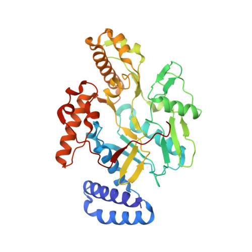
    5G65, 5G66, 5G67, 5G68, 5G69, 5G6A, 5G6B, 5G6C, 5G6D, 5G6E, 5G6F, 5G6G, 5G6H, 5G6I, 5G6J, 5G6K, 5G6L, 5G6M, 5G6N, 5G6O, 5G6P, 5G6Q

  • PubMed Abstract: 

    Nitric oxide is produced in Gram-positive pathogens Bacillus anthracis and Staphylococcus aureus by the bacterial isoform of nitric oxide synthase (NOS). Inhibition of bacterial nitric oxide synthase (bNOS) has been identified as a promising antibacterial strategy for targeting methicillin-resistant S. aureus [Holden, J. K., et al. (2015) Chem. Biol. 22, 785-779]. One class of NOS inhibitors that demonstrates antimicrobial efficacy utilizes an aminoquinoline scaffold. Here we report on a variety of aminoquinolines that target the bacterial NOS active site, in part, by binding to a hydrophobic patch that is unique to bNOS. Through mutagenesis and crystallographic studies, our findings demonstrate that aminoquinolines are an excellent scaffold for further aiding in the development of bNOS specific inhibitors.


  • Organizational Affiliation
    • Department of Molecular Biology and Biochemistry, ‡Department of Pharmaceutical Sciences, and §Department of Chemistry, University of California , Irvine, California 92697-3900, United States.

Macromolecule Content 

  • Total Structure Weight: 43.14 kDa 
  • Atom Count: 3,284 
  • Modeled Residue Count: 362 
  • Deposited Residue Count: 363 
  • Unique protein chains: 1

Macromolecules

Find similar proteins by:|  3D Structure
Entity ID: 1
MoleculeChains  Sequence LengthOrganismDetailsImage
NITRIC OXIDE SYNTHASE OXYGENASE363Bacillus subtilis subsp. subtilis str. 168Mutation(s): 3 
EC: 1.14.13.165 (PDB Primary Data), 1.14.14.47 (UniProt)
UniProt
Find proteins for O34453 (Bacillus subtilis (strain 168))
Explore O34453 
Go to UniProtKB:  O34453
Entity Groups
Sequence Clusters30% Identity50% Identity70% Identity90% Identity95% Identity100% Identity
UniProt GroupO34453
Sequence Annotations
Expand
Reference Sequence

Small Molecules

Ligands 5 Unique
IDChains Name / Formula / InChI Key2D Diagram3D Interactions
HEM

Query on HEM


Download:Ideal Coordinates CCD File
B [auth A]PROTOPORPHYRIN IX CONTAINING FE
C34 H32 Fe N4 O4
KABFMIBPWCXCRK-RGGAHWMASA-L
M85

Query on M85


Download:Ideal Coordinates CCD File
E [auth A]7-[[3-(methylaminomethyl)phenoxy]methyl]quinolin-2-amine
C18 H19 N3 O
QFZGTQOWVVISDX-UHFFFAOYSA-N
H4B

Query on H4B


Download:Ideal Coordinates CCD File
C [auth A]5,6,7,8-TETRAHYDROBIOPTERIN
C9 H15 N5 O3
FNKQXYHWGSIFBK-RPDRRWSUSA-N
GOL

Query on GOL


Download:Ideal Coordinates CCD File
F [auth A],
G [auth A]
GLYCEROL
C3 H8 O3
PEDCQBHIVMGVHV-UHFFFAOYSA-N
CL

Query on CL


Download:Ideal Coordinates CCD File
D [auth A]CHLORIDE ION
Cl
VEXZGXHMUGYJMC-UHFFFAOYSA-M

Experimental Data & Validation

Experimental Data

  • Method: X-RAY DIFFRACTION
  • Resolution: 1.98 Å
  • R-Value Free:  0.217 (Depositor), 0.218 (DCC) 
  • R-Value Work:  0.167 (Depositor), 0.167 (DCC) 
  • R-Value Observed: 0.169 (Depositor) 
Space Group: P 21 21 2
Unit Cell:
Length ( Å )Angle ( ˚ )
a = 80.67α = 90
b = 94.49β = 90
c = 61.91γ = 90
Software Package:
Software NamePurpose
PHENIXrefinement
MOSFLMdata reduction
Aimlessdata scaling

Structure Validation

View Full Validation Report


Ligand Structure Quality Assessment 


Entry History 

Deposition Data

Revision History  (Full details and data files)

  • Version 1.0: 2016-09-21
    Type: Initial release
  • Version 1.1: 2016-10-19
    Changes: Database references
  • Version 1.2: 2019-01-30
    Changes: Data collection, Experimental preparation, Other
  • Version 1.3: 2019-02-06
    Changes: Data collection, Database references, Experimental preparation
  • Version 1.4: 2024-01-10
    Changes: Data collection, Database references, Derived calculations, Other, Refinement description