5G4K | pdb_00005g4k

Phloroglucinol reductase from Clostridium sp. apo-form


Experimental Data Snapshot

  • Method: X-RAY DIFFRACTION
  • Resolution: 1.74 Å
  • R-Value Free: 
    0.182 (Depositor) 
  • R-Value Work: 
    0.157 (Depositor) 
  • R-Value Observed: 
    0.158 (Depositor) 

Starting Model: experimental
View more details

wwPDB Validation 3D Report Full Report

Validation slider image for 5G4K

This is version 1.2 of the entry. See complete history

Literature

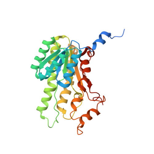
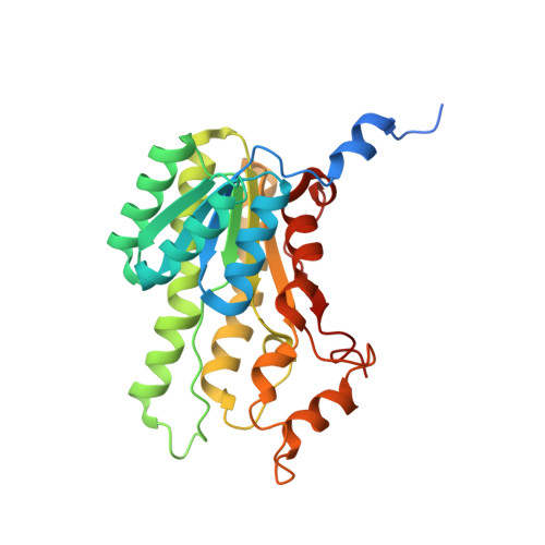
Biocatalytic Properties and Structural Analysis of Phloroglucinol Reductases.

Conradt, D.Hermann, B.Gerhardt, S.Einsle, O.Mueller, M.

(2016) Angew Chem Int Ed Engl 55: 15531

  • DOI: https://doi.org/10.1002/anie.201607494
  • Primary Citation Related Structures: 
    5G4K, 5G4L

  • PubMed Abstract: 

    Phloroglucinol reductases (PGRs) are involved in anaerobic degradation in bacteria, in which they catalyze the dearomatization of phloroglucinol into dihydrophloroglucinol. We identified three PGRs, from different bacterial species, that are members of the family of NAD(P)H-dependent short-chain dehydrogenases/reductases (SDRs). In addition to catalyzing the reduction of the physiological substrate, the three enzymes exhibit activity towards 2,4,6-trihydroxybenzaldehyde, 2,4,6-trihydroxyacetophenone, and methyl 2,4,6-trihydroxybenzoate. Structural elucidation of PGRcl and comparison to known SDRs revealed a high degree of conservation. Several amino acid positions were identified as being conserved within the PGR subfamily and might be involved in substrate differentiation. The results enable the enzymatic dearomatization of monoaromatic phenol derivatives and provide insight into the functional diversity that may be found in families of enzymes displaying a high degree of structural homology.


  • Organizational Affiliation
    • Institut für Pharmazeutische Wissenschaften, Albert-Ludwigs-Universität Freiburg, Albertstrasse 25, 79104, Freiburg, Germany.

Macromolecule Content 

  • Total Structure Weight: 62.27 kDa 
  • Atom Count: 4,356 
  • Modeled Residue Count: 528 
  • Deposited Residue Count: 572 
  • Unique protein chains: 2

Macromolecules

Find similar proteins by:|  3D Structure
Entity ID: 1
MoleculeChains  Sequence LengthOrganismDetailsImage
OXIDOREDUCTASE, SHORT CHAIN DEHYDROGENASE/REDUCTASE FAMILY PROTEIN286Clostridium sp.Mutation(s): 0 
Find similar proteins by:|  3D Structure
Entity ID: 2
MoleculeChains  Sequence LengthOrganismDetailsImage
OXIDOREDUCTASE, SHORT CHAIN DEHYDROGENASE/REDUCTASE FAMILY PROTEIN286Clostridium sp.Mutation(s): 0 

Experimental Data & Validation

Experimental Data

  • Method: X-RAY DIFFRACTION
  • Resolution: 1.74 Å
  • R-Value Free:  0.182 (Depositor) 
  • R-Value Work:  0.157 (Depositor) 
  • R-Value Observed: 0.158 (Depositor) 
Space Group: P 21 21 2
Unit Cell:
Length ( Å )Angle ( ˚ )
a = 89.756α = 90
b = 125.391β = 90
c = 55.468γ = 90
Software Package:
Software NamePurpose
BUSTERrefinement
autoPROCdata reduction
Aimlessdata scaling
MOLREPphasing

Structure Validation

View Full Validation Report


Entry History 

Deposition Data

Revision History  (Full details and data files)

  • Version 1.0: 2016-12-07
    Type: Initial release
  • Version 1.1: 2016-12-14
    Changes: Database references
  • Version 1.2: 2024-01-10
    Changes: Data collection, Database references, Other, Refinement description