5G4H | pdb_00005g4h

1.50 A resolution catechol (1,2-dihydroxybenzene) inhibited Sporosarcina pasteurii urease


Experimental Data Snapshot

  • Method: X-RAY DIFFRACTION
  • Resolution: 1.50 Å
  • R-Value Free: 
    0.154 (Depositor), 0.168 (DCC) 
  • R-Value Work: 
    0.121 (Depositor), 0.139 (DCC) 
  • R-Value Observed: 
    0.123 (Depositor) 

Starting Model: experimental
View more details

wwPDB Validation 3D Report Full Report

Validation slider image for 5G4H

This is version 1.2 of the entry. See complete history

Literature

Inactivation of Urease by Catechol: Kinetics and Structure.

Mazzei, L.Cianci, M.Musiani, F.Lente, G.Palombo, M.Ciurli, S.

(2016) J Inorg Biochem 166: 182

  • DOI: https://doi.org/10.1016/j.jinorgbio.2016.11.016
  • Primary Citation Related Structures: 
    5G4H

  • PubMed Abstract: 

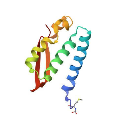
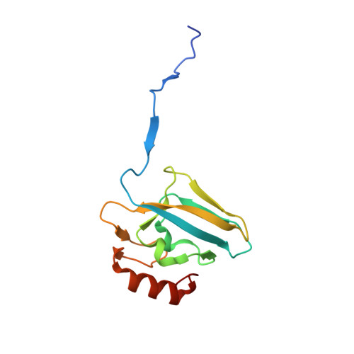
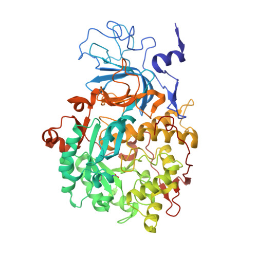
    Urease is a Ni(II)-containing enzyme that catalyzes the hydrolysis of urea to yield ammonia and carbamate at a rate 10 15 times higher than the uncatalyzed reaction. Urease is a virulence factor of several human pathogens, in addition to decreasing the efficiency of soil organic nitrogen fertilization. Therefore, efficient urease inhibitors are actively sought. In this study, we describe a molecular characterization of the interaction between urease from Sporosarcina pasteurii (SPU) and Canavalia ensiformis (jack bean, JBU) with catechol, a model polyphenol. In particular, catechol irreversibly inactivates both SPU and JBU with a complex radical-based autocatalytic multistep mechanism. The crystal structure of the SPU-catechol complex, determined at 1.50Å resolution, reveals the structural details of the enzyme inhibition.


  • Organizational Affiliation
    • Laboratory of Bioinorganic Chemistry, Department of Pharmacy and Biotechnology, University of Bologna, Italy.

Macromolecule Content 

  • Total Structure Weight: 88.44 kDa 
  • Atom Count: 7,020 
  • Modeled Residue Count: 792 
  • Deposited Residue Count: 796 
  • Unique protein chains: 3

Macromolecules

Find similar proteins by:|  3D Structure
Entity ID: 1
MoleculeChains  Sequence LengthOrganismDetailsImage
UREASE SUBUNIT GAMMA100Sporosarcina pasteuriiMutation(s): 0 
EC: 3.5.1.5
UniProt
Find proteins for P41022 (Sporosarcina pasteurii)
Explore P41022 
Go to UniProtKB:  P41022
Entity Groups
Sequence Clusters30% Identity50% Identity70% Identity90% Identity95% Identity100% Identity
UniProt GroupP41022
Sequence Annotations
Expand
Reference Sequence
Find similar proteins by:|  3D Structure
Entity ID: 2
MoleculeChains  Sequence LengthOrganismDetailsImage
UREASE SUBUNIT BETA126Sporosarcina pasteuriiMutation(s): 0 
EC: 3.5.1.5
UniProt
Find proteins for P41021 (Sporosarcina pasteurii)
Explore P41021 
Go to UniProtKB:  P41021
Entity Groups
Sequence Clusters30% Identity50% Identity70% Identity90% Identity95% Identity100% Identity
UniProt GroupP41021
Sequence Annotations
Expand
Reference Sequence
Find similar proteins by:|  3D Structure
Entity ID: 3
MoleculeChains  Sequence LengthOrganismDetailsImage
UREASE SUBUNIT ALPHA570Sporosarcina pasteuriiMutation(s): 0 
EC: 3.5.1.5

Small Molecules

Ligands 5 Unique
IDChains Name / Formula / InChI Key2D Diagram3D Interactions
CAQ

Query on CAQ


Download:Ideal Coordinates CCD File
L [auth C]CATECHOL
C6 H6 O2
YCIMNLLNPGFGHC-UHFFFAOYSA-N
SO4

Query on SO4


Download:Ideal Coordinates CCD File
BA [auth C]
D [auth A]
G [auth B]
I [auth B]
M [auth C]
BA [auth C],
D [auth A],
G [auth B],
I [auth B],
M [auth C],
O [auth C]
SULFATE ION
O4 S
QAOWNCQODCNURD-UHFFFAOYSA-L
EDO

Query on EDO


Download:Ideal Coordinates CCD File
AA [auth C]
E [auth A]
F [auth A]
H [auth B]
P [auth C]
AA [auth C],
E [auth A],
F [auth A],
H [auth B],
P [auth C],
Q [auth C],
R [auth C],
S [auth C],
T [auth C],
U [auth C],
V [auth C],
W [auth C],
X [auth C],
Y [auth C],
Z [auth C]
1,2-ETHANEDIOL
C2 H6 O2
LYCAIKOWRPUZTN-UHFFFAOYSA-N
NI

Query on NI


Download:Ideal Coordinates CCD File
J [auth C],
K [auth C]
NICKEL (II) ION
Ni
VEQPNABPJHWNSG-UHFFFAOYSA-N
OH

Query on OH


Download:Ideal Coordinates CCD File
N [auth C]HYDROXIDE ION
H O
XLYOFNOQVPJJNP-UHFFFAOYSA-M
Modified Residues  2 Unique
IDChains TypeFormula2D DiagramParent
CXM
Query on CXM
A
L-PEPTIDE LINKINGC6 H11 N O4 SMET
KCX
Query on KCX
C
L-PEPTIDE LINKINGC7 H14 N2 O4LYS

Experimental Data & Validation

Experimental Data

  • Method: X-RAY DIFFRACTION
  • Resolution: 1.50 Å
  • R-Value Free:  0.154 (Depositor), 0.168 (DCC) 
  • R-Value Work:  0.121 (Depositor), 0.139 (DCC) 
  • R-Value Observed: 0.123 (Depositor) 
Space Group: P 63 2 2
Unit Cell:
Length ( Å )Angle ( ˚ )
a = 131.276α = 90
b = 131.276β = 90
c = 189.017γ = 120
Software Package:
Software NamePurpose
REFMACrefinement
XDSdata reduction
Aimlessdata scaling
CCP4phasing

Structure Validation

View Full Validation Report


Entry History 

Deposition Data

Revision History  (Full details and data files)

  • Version 1.0: 2016-12-07
    Type: Initial release
  • Version 1.1: 2018-03-07
    Changes: Data collection
  • Version 1.2: 2024-01-10
    Changes: Data collection, Database references, Derived calculations, Other, Refinement description