5G35 | pdb_00005g35

Structure of Rad14 in complex with acetylaminopyren-C8-guanine containing DNA


Experimental Data Snapshot

  • Method: X-RAY DIFFRACTION
  • Resolution: 2.00 Å
  • R-Value Free: 
    0.254 (Depositor), 0.260 (DCC) 
  • R-Value Work: 
    0.217 (Depositor), 0.227 (DCC) 
  • R-Value Observed: 
    0.219 (Depositor) 

Starting Model: experimental
View more details

wwPDB Validation 3D Report Full Report

Validation slider image for 5G35

This is version 2.1 of the entry. See complete history

Literature

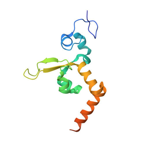
Structural Basis for Bulky Adduct DNA Lesion Recognition by the Nucleotide Excision Repair Protein Rad14.

Schneider, S.Simon, N.Ebert, C.

(2016) Chemistry 22: 10782

  • DOI: https://doi.org/10.1002/chem.201602438
  • Primary Citation Related Structures: 
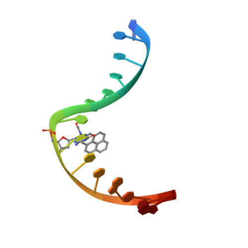
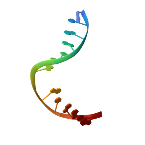
    5G32, 5G33, 5G34, 5G35

  • PubMed Abstract: 

    Heterocyclic aromatic amines react with purine bases and result in bulky DNA adducts that cause mutations. Such structurally diverse lesions are substrates for the nucleotide excision repair (NER). It is thought that the NER machinery recognises and verifies distorted DNA conformations, also involving the xeroderma pigmentosum group A and C proteins (XPA, XPC) that act as a scaffold between the DNA substrate and several other NER proteins. Here we present the synthesis of DNA molecules containing the polycyclic, aromatic amine C8-guanine lesions acetylaminophenyl, acetylaminonaphthyl, acetylaminoanthryl, and acetylaminopyrenyl, as well as their crystal structures in complex with the yeast XPA homologue Rad14. This work further substantiates the indirect lesion-detection mechanism employed by the NER system that recognises destabilised and deformable DNA structures.


  • Organizational Affiliation
    • Center for Integrated Protein Science Munich CIPSM, Department of Chemistry, Ludwig-Maximilians Universität München, Butenandtstrasse 13, 81377, München, Germany.

Macromolecule Content 

  • Total Structure Weight: 49.65 kDa 
  • Atom Count: 3,093 
  • Modeled Residue Count: 285 
  • Deposited Residue Count: 320 
  • Unique protein chains: 1
  • Unique nucleic acid chains: 2

Macromolecules


Find similar proteins by:|  3D Structure
Entity ID: 1
MoleculeChains  Sequence LengthOrganismDetailsImage
RAD14
A, B
131Saccharomyces cerevisiaeMutation(s): 0 
Gene Names: RAD14YMR201CYM8325.02C
UniProt
Find proteins for P28519 (Saccharomyces cerevisiae (strain ATCC 204508 / S288c))
Explore P28519 
Go to UniProtKB:  P28519
Entity Groups
Sequence Clusters30% Identity50% Identity70% Identity90% Identity95% Identity100% Identity
UniProt GroupP28519
Sequence Annotations
Expand
Reference Sequence
Find similar nucleic acids by:  Sequence
Entity ID: 2
MoleculeChains LengthOrganismImage
5'-D(*GP*CP*TP*CP*TP*AP*8PYP*TP*CP*AP*TP*CP*AP*CP)-3'
C, E
15synthetic construct
Sequence Annotations
Expand
Reference Sequence
Find similar nucleic acids by:  Sequence
Entity ID: 3
MoleculeChains LengthOrganismImage
5'-D(*GP*TP*GP*AP*TP*GP*AP*CP*GP*TP*AP*GP*AP*GP)-3'
D, F
14synthetic construct
Sequence Annotations
Expand
Reference Sequence

Experimental Data & Validation

Experimental Data

  • Method: X-RAY DIFFRACTION
  • Resolution: 2.00 Å
  • R-Value Free:  0.254 (Depositor), 0.260 (DCC) 
  • R-Value Work:  0.217 (Depositor), 0.227 (DCC) 
  • R-Value Observed: 0.219 (Depositor) 
Space Group: P 41
Unit Cell:
Length ( Å )Angle ( ˚ )
a = 53.17α = 90
b = 53.17β = 90
c = 131.307γ = 90
Software Package:
Software NamePurpose
REFMACrefinement
XDSdata reduction
REFMACphasing

Structure Validation

View Full Validation Report


Entry History 

Deposition Data

Revision History  (Full details and data files)

  • Version 1.0: 2016-06-01
    Type: Initial release
  • Version 1.1: 2016-06-08
    Changes: Database references
  • Version 1.2: 2016-08-03
    Changes: Database references
  • Version 2.0: 2019-05-15
    Changes: Data collection, Derived calculations, Other, Polymer sequence
  • Version 2.1: 2024-01-10
    Changes: Data collection, Database references, Derived calculations, Other, Refinement description