5FYD | pdb_00005fyd

Structural and biochemical insights into 7beta-hydroxysteroid dehydrogenase stereoselectivity


Experimental Data Snapshot

  • Method: X-RAY DIFFRACTION
  • Resolution: 1.60 Å
  • R-Value Free: 
    0.209 (Depositor), 0.217 (DCC) 
  • R-Value Work: 
    0.176 (Depositor), 0.187 (DCC) 
  • R-Value Observed: 
    0.178 (Depositor) 

Starting Model: experimental
View more details

wwPDB Validation 3D Report Full Report

Validation slider image for 5FYD

This is version 1.2 of the entry. See complete history

Literature

Structural and Biochemical Insights Into 7Beta-Hydroxysteroid Dehydrogenase Stereoselectivity.

Savino, S.Ferrandi, E.E.Forneris, F.Rovida, S.Riva, S.Monti, D.Mattevi, A.

(2016) Proteins 84: 859

  • DOI: https://doi.org/10.1002/prot.25036
  • Primary Citation Related Structures: 
    5FYD

  • PubMed Abstract: 

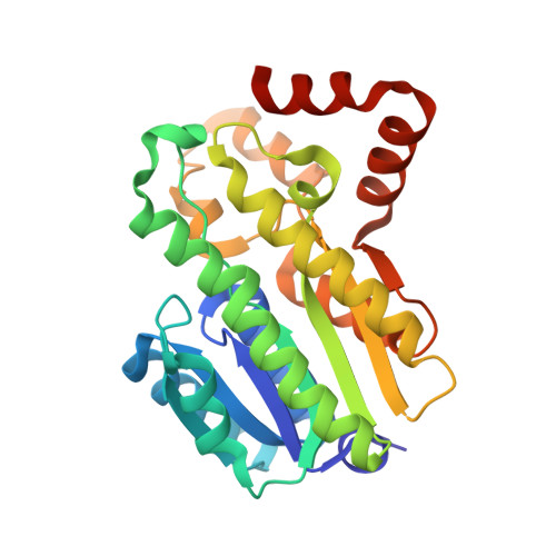
    Hydroxysteroid dehydrogenases are of great interest as biocatalysts for transformations involving steroid substrates. They feature a high degree of stereo- and regio-selectivity, acting on a defined atom with a specific configuration of the steroid nucleus. The crystal structure of 7β-hydroxysteroid dehydrogenase from Collinsella aerofaciens reveals a loop gating active-site accessibility, the bases of the specificity for NADP(+) , and the general architecture of the steroid binding site. Comparison with 7α-hydroxysteroid dehydrogenase provides a rationale for the opposite stereoselectivity. The presence of a C-terminal extension reshapes the substrate site of the β-selective enzyme, possibly leading to an inverted orientation of the bound substrate. Proteins 2016; 84:859-865. © 2016 Wiley Periodicals, Inc.


  • Organizational Affiliation
    • Department of Biology and Biotechnology, University of Pavia, via Ferrata 9, Pavia, 27100, Italy.

Macromolecule Content 

  • Total Structure Weight: 57.66 kDa 
  • Atom Count: 4,269 
  • Modeled Residue Count: 511 
  • Deposited Residue Count: 526 
  • Unique protein chains: 1

Macromolecules

Find similar proteins by:|  3D Structure
Entity ID: 1
MoleculeChains  Sequence LengthOrganismDetailsImage
OXIDOREDUCTASE, SHORT CHAIN DEHYDROGENASE/REDUCTASE FAMILY PROTEIN
A, B
263Collinsella aerofaciensMutation(s): 0 
EC: 1.1.1.201
UniProt
Find proteins for A4ECA9 (Collinsella aerofaciens (strain ATCC 25986 / DSM 3979 / JCM 10188 / KCTC 3647 / NCTC 11838 / VPI 1003))
Explore A4ECA9 
Go to UniProtKB:  A4ECA9
Entity Groups
Sequence Clusters30% Identity50% Identity70% Identity90% Identity95% Identity100% Identity
UniProt GroupA4ECA9
Sequence Annotations
Expand
Reference Sequence

Small Molecules

Ligands 1 Unique
IDChains Name / Formula / InChI Key2D Diagram3D Interactions
GOL

Query on GOL


Download:Ideal Coordinates CCD File
C [auth A]GLYCEROL
C3 H8 O3
PEDCQBHIVMGVHV-UHFFFAOYSA-N

Experimental Data & Validation

Experimental Data

  • Method: X-RAY DIFFRACTION
  • Resolution: 1.60 Å
  • R-Value Free:  0.209 (Depositor), 0.217 (DCC) 
  • R-Value Work:  0.176 (Depositor), 0.187 (DCC) 
  • R-Value Observed: 0.178 (Depositor) 
Space Group: P 21 21 2
Unit Cell:
Length ( Å )Angle ( ˚ )
a = 114.77α = 90
b = 83.04β = 90
c = 47.91γ = 90
Software Package:
Software NamePurpose
REFMACrefinement
iMOSFLMdata reduction
Aimlessdata scaling
PHASERphasing

Structure Validation

View Full Validation Report


Entry History 

Deposition Data

Revision History  (Full details and data files)

  • Version 1.0: 2016-04-06
    Type: Initial release
  • Version 1.1: 2016-06-01
    Changes: Database references
  • Version 1.2: 2024-01-10
    Changes: Data collection, Database references, Derived calculations, Other, Refinement description