5FXE | pdb_00005fxe

Crystal structure of eugenol oxidase in complex with coniferyl alcohol


Experimental Data Snapshot

  • Method: X-RAY DIFFRACTION
  • Resolution: 1.90 Å
  • R-Value Free: 
    0.211 (Depositor), 0.221 (DCC) 
  • R-Value Work: 
    0.161 (Depositor), 0.172 (DCC) 
  • R-Value Observed: 
    0.164 (Depositor) 

Starting Model: experimental
View more details

wwPDB Validation 3D Report Full Report

Validation slider image for 5FXE

Ligand Structure Quality Assessment 


This is version 1.2 of the entry. See complete history

Literature

Biocatalytic Properties and Structural Analysis of Eugenol Oxidase from Rhodococcus Jostii Rha1: A Versatile Oxidative Biocatalyst.

Nguyen, Q.De Gonzalo, G.Binda, C.Rioz-Martinez, A.Mattevi, A.Fraaije, M.W.

(2016) Chembiochem 17: 1359

  • DOI: https://doi.org/10.1002/cbic.201600148
  • Primary Citation Related Structures: 
    5FXD, 5FXE, 5FXF, 5FXP

  • PubMed Abstract: 

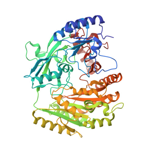
    Eugenol oxidase (EUGO) from Rhodococcus jostii RHA1 had previously been shown to convert only a limited set of phenolic compounds. In this study, we have explored the biocatalytic potential of this flavoprotein oxidase, resulting in a broadened substrate scope and a deeper insight into its structural properties. In addition to the oxidation of vanillyl alcohol and the hydroxylation of eugenol, EUGO can efficiently catalyze the dehydrogenation of various phenolic ketones and the selective oxidation of a racemic secondary alcohol-4-(1-hydroxyethyl)-2-methoxyphenol. EUGO was also found to perform the kinetic resolution of a racemic secondary alcohol. Crystal structures of the enzyme in complexes with isoeugenol, coniferyl alcohol, vanillin, and benzoate have been determined. The catalytic center is a remarkable solvent-inaccessible cavity on the si side of the flavin cofactor. Structural comparison with vanillyl alcohol oxidase from Penicillium simplicissimum highlights a few localized changes that correlate with the selectivity of EUGO for phenolic substrates bearing relatively small p-substituents while tolerating o-methoxy substituents.


  • Organizational Affiliation
    • Molecular Enzymology, Groningen Biomolecular Sciences and, Biotechnology Institute, University of Groningen, Nijenborgh 4, 9747 AG, Groningen, NL.

Macromolecule Content 

  • Total Structure Weight: 119.96 kDa 
  • Atom Count: 8,891 
  • Modeled Residue Count: 1,050 
  • Deposited Residue Count: 1,052 
  • Unique protein chains: 1

Macromolecules

Find similar proteins by:|  3D Structure
Entity ID: 1
MoleculeChains  Sequence LengthOrganismDetailsImage
EUGENOL OXIDASE
A, B
526Rhodococcus jostii RHA1Mutation(s): 0 
EC: 1.1.3.38
UniProt
Find proteins for Q0SBK1 (Rhodococcus jostii (strain RHA1))
Explore Q0SBK1 
Go to UniProtKB:  Q0SBK1
Entity Groups
Sequence Clusters30% Identity50% Identity70% Identity90% Identity95% Identity100% Identity
UniProt GroupQ0SBK1
Sequence Annotations
Expand
Reference Sequence

Small Molecules

Ligands 3 Unique
IDChains Name / Formula / InChI Key2D Diagram3D Interactions
FAD

Query on FAD


Download:Ideal Coordinates CCD File
C [auth A],
I [auth B]
FLAVIN-ADENINE DINUCLEOTIDE
C27 H33 N9 O15 P2
VWWQXMAJTJZDQX-UYBVJOGSSA-N
CIY

Query on CIY


Download:Ideal Coordinates CCD File
D [auth A],
J [auth B]
(2E)-3-(4-hydroxy-3-methoxyphenyl)prop-2-enal
C10 H10 O3
DKZBBWMURDFHNE-NSCUHMNNSA-N
GOL

Query on GOL


Download:Ideal Coordinates CCD File
E [auth A]
F [auth A]
G [auth A]
H [auth A]
K [auth B]
E [auth A],
F [auth A],
G [auth A],
H [auth A],
K [auth B],
L [auth B]
GLYCEROL
C3 H8 O3
PEDCQBHIVMGVHV-UHFFFAOYSA-N

Experimental Data & Validation

Experimental Data

  • Method: X-RAY DIFFRACTION
  • Resolution: 1.90 Å
  • R-Value Free:  0.211 (Depositor), 0.221 (DCC) 
  • R-Value Work:  0.161 (Depositor), 0.172 (DCC) 
  • R-Value Observed: 0.164 (Depositor) 
Space Group: P 21 21 21
Unit Cell:
Length ( Å )Angle ( ˚ )
a = 57.47α = 90
b = 97.17β = 90
c = 179.05γ = 90
Software Package:
Software NamePurpose
REFMACrefinement
MOSFLMdata reduction
Aimlessdata scaling
MOLREPphasing

Structure Validation

View Full Validation Report


Ligand Structure Quality Assessment 


Entry History 

Deposition Data

Revision History  (Full details and data files)

  • Version 1.0: 2016-07-27
    Type: Initial release
  • Version 1.1: 2024-01-10
    Changes: Data collection, Database references, Derived calculations, Other, Refinement description
  • Version 1.2: 2024-10-09
    Changes: Structure summary