5FW9 | pdb_00005fw9

Human Spectrin SH3 domain D48G, E7Y, K60Y


Experimental Data Snapshot

  • Method: X-RAY DIFFRACTION
  • Resolution: 1.55 Å
  • R-Value Free: 
    0.249 (Depositor), 0.243 (DCC) 
  • R-Value Work: 
    0.203 (Depositor), 0.198 (DCC) 
  • R-Value Observed: 
    0.205 (Depositor) 

Starting Model: experimental
View more details

wwPDB Validation 3D Report Full Report

Validation slider image for 5FW9

This is version 1.1 of the entry. See complete history

Literature

Human Spectrin SH3 Domain D48G, E7Y, K60Y

Navarro, S.Gallego, P.Diaz, M.Ventura, S.Reverter, D.

To be published.

Macromolecule Content 

  • Total Structure Weight: 7.24 kDa 
  • Atom Count: 545 
  • Modeled Residue Count: 57 
  • Deposited Residue Count: 62 
  • Unique protein chains: 1

Macromolecules

Find similar proteins by:|  3D Structure
Entity ID: 1
MoleculeChains  Sequence LengthOrganismDetailsImage
SPECTRIN ALPHA CHAIN, NON-ERYTHROCYTIC 162Homo sapiensMutation(s): 3 
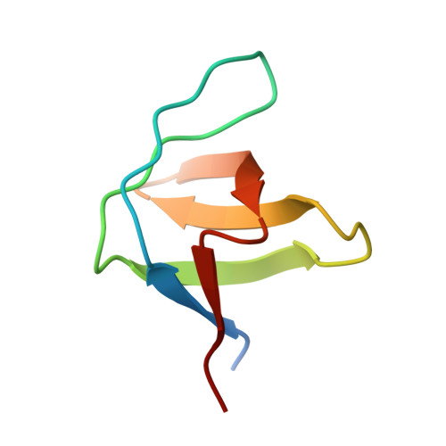
UniProt & NIH Common Fund Data Resources
Find proteins for Q13813 (Homo sapiens)
Explore Q13813 
Go to UniProtKB:  Q13813
PHAROS:  Q13813
GTEx:  ENSG00000197694 
Entity Groups
Sequence Clusters30% Identity50% Identity70% Identity90% Identity95% Identity100% Identity
UniProt GroupQ13813
Sequence Annotations
Expand
Reference Sequence

Experimental Data & Validation

Experimental Data

  • Method: X-RAY DIFFRACTION
  • Resolution: 1.55 Å
  • R-Value Free:  0.249 (Depositor), 0.243 (DCC) 
  • R-Value Work:  0.203 (Depositor), 0.198 (DCC) 
  • R-Value Observed: 0.205 (Depositor) 
Space Group: P 21 21 21
Unit Cell:
Length ( Å )Angle ( ˚ )
a = 33.234α = 90
b = 42.005β = 90
c = 50.336γ = 90
Software Package:
Software NamePurpose
REFMACrefinement

Structure Validation

View Full Validation Report


Entry History 

Deposition Data

Revision History  (Full details and data files)

  • Version 1.0: 2016-12-28
    Type: Initial release
  • Version 1.1: 2024-01-10
    Changes: Data collection, Database references, Other, Refinement description